tips-lateral-flow-readouts-crispr-cas-strategies

04/30
2020

Lateral Flow Readout for CRISPR/Cas-based detection strategies

Test– and Control-line of the HybriDetect

SHERLOCK and DETECTR are combinations of isothermal amplification and sensitive CRISPR/Cas-based amplicon recognition. The compatibility with the rapid, simple and equipment-free HybriDetect lateral flow platform is an essential feature of these methods. Interpretation of the lateral flow strips is very intuitive and easy.

The way HybriDetect is used for CRISPR applications can lead to confusions about the Test- and the Control-line. The following figure explains this difference.

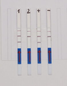
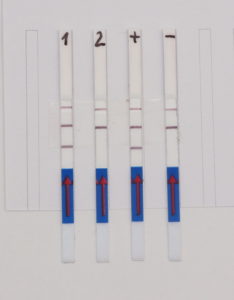
As shown in Figure 1, the interpretation of the test strips can vary using different detection strategies. Compared to the HybriDetect instructions for use, the T- and C-lines are swapped in CRSIPR/Cas-associated detection strategies. This leads to a completely different assessment of the resulting signals. The following article is intended to explain this discrepancy in detail and it provides some important information on how to use the HybriDetect in combination with the SHERLOCK / DETECTR method.

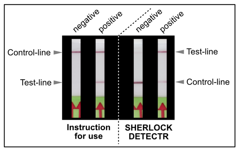
Figure 1. Difference of test result interpretation between the HybriDetect Instructions for use and the SHERLOCK-/DETECTR-protocol guidelines

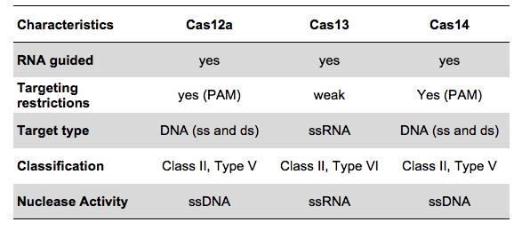
What defines the control- and test-lines?
The “T and the “C” in Lateral Flow Devices (LFDs)

The presence of at least one visually detectable test-line (T-line) is the essential feature of LFDs. In most cases the presence of the T-line correlates with a positive test result. Additionally, a control-line (C-line) confirms the general functionality of LFA. This is absolutely necessary for differentiation between valid negative results and invalid, non-interpretable test results.

The HybriDetect design is basically comparable to the structure of a classic LFD, but the idea behind this detection platform differs.

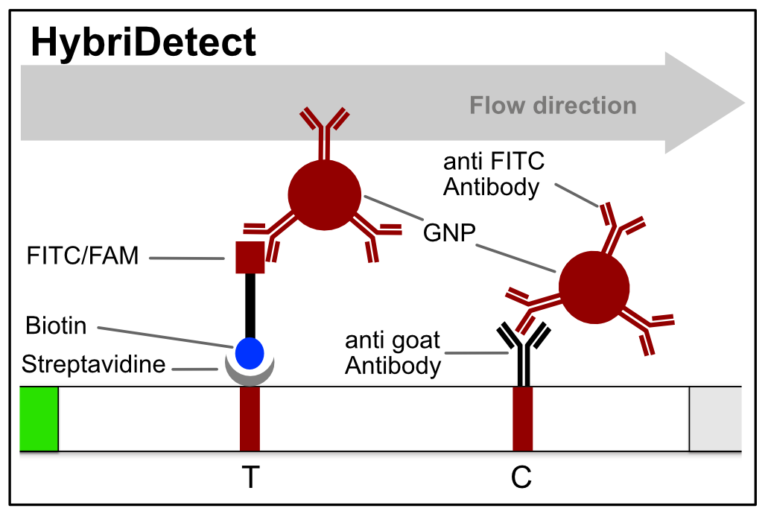
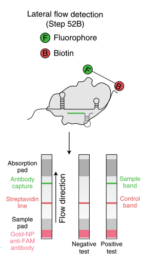
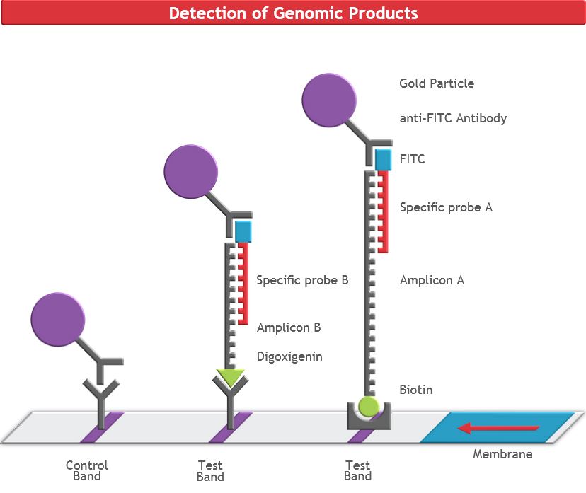
Figure 2. Composition of the HybriDetect. Biotin and FITC/FAM are introduced into LFA through sample application. The control-line (C) will show up, if you run the HybriDetect without sample. GNP – Goldnanoparticle

HybriDetect and signal generation strategies

The idea behind the HybriDetect platform differs fundamentally from “classic” LFAs. The HybriDetect and HybriDetect 2T are universal test strips, developed to work in multiple detection concepts and not in just one setting. They can be used to detect amplicons, proteins, ribosomal RNAs, whole cells or metabolites. Over the years various detection strategies have been developed, in which the HybriDetect is used.

In most constellations, biotin and FITC are somehow incorporated into analytes or reaction products, which results in the formation of the predefined test-line(s). Popular applications combined with HybriDetect readout are especially DNA amplification techniques such as PCR, RPA or LAMP. However, these LFA-compatible methods have one feature in common: they are designed according to the described Label Incorporation Strategy.

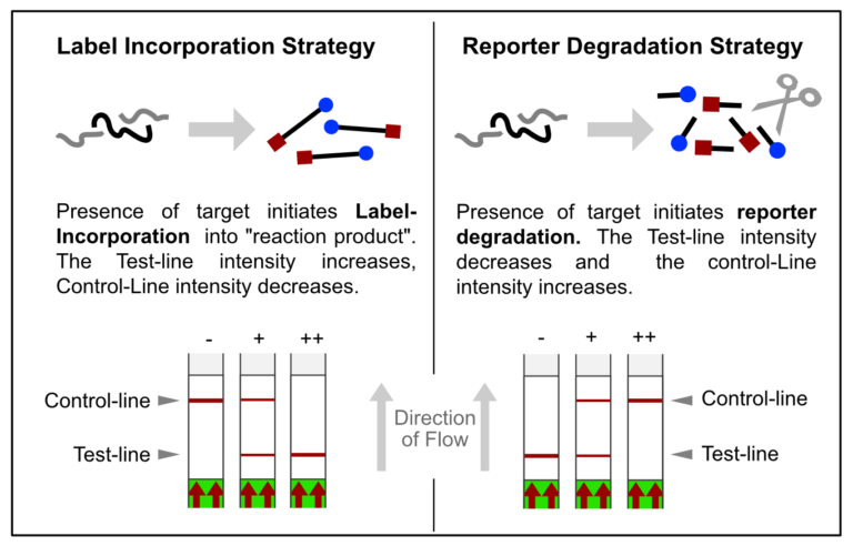
Figure 3. Overview of two LFA-related signal generation stratefies. Label incorporation strategy leads to Test-line formation, whereas reporter degradation will end up in decreased T-line and increased C-line intensity.

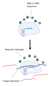
In contrast to this, methods such as SHERLOCK or DETECTR generate amplification products, which do not directly contribute to signal generation. Rather, CRISPR complexes are able to specifically and sensitively recognize defined amplicon-related sequences. Successful recognition leads to the activation of the Cas-protein associated collateral nuclease activity. The ssRNA- / ssDNA- / dsDNA – degrading activity is the basis for signal generation in LFA. This is achieved by using the reporter degradation strategy.
The inverse approach – degradation of the “original” T-line

The key to the functionality of a CRISPR-associated nuclease assay combined with a lateral flow readout is the use of a so-called reporter. Such reporters are short single-stranded DNA- or RNA-Oligos with terminal biotin- and FITC-/ FAM-modifications. Reporter sequences are chosen according to cleavage preferences of the used Cas-protein. The following table shows a brief overview of some reporters, which are used in the most common CRISPR/Cas-based detection systems.

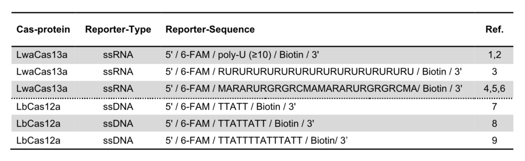
Table 1. Overview of reporter sequences used in CRISPR/Cas-based detection methods combined with Lateral Flow readout

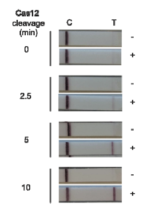
An intense test-line appears if a defined amount of reporter is analyzed with the HybriDetect. But if reporter degradation is initiated due to the onset of nuclease activity, the intensity of the T-line consequently decreases.

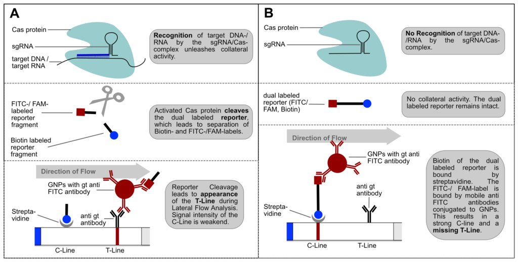
If the reporter is used in a very defined concentration, the majority of the gold conjugate is trapped at the T-line, which leads to an almost complete extinction of the C-line. It is precisely this effect, that enables the T- and C-line swap for CRISPR/Cas-associated detection methods. Ultimately, it’s just a matter of definition and there is no change in HybriDetect composition! The following figure explains the CRISPR/Cas-based detection strategy with newly defined T- and C-line.

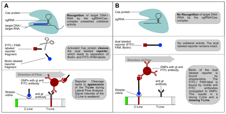
Figure 4. Switching C- and T-line: General mechanism of CRISPR/Cas-mediated detection of nucleic acids via HybriDetect Lateral Flow. (A) The presence of the genetic target leads to a positive test result. (B) The absence of the specific genetic target leads to a negative test result.

Switching test- and control-line has two major advantages: An increasing or appearing signal correlates with a positive test result. Negative results are interpreted if no signal appears. This positive correlation is much more logical for most people and avoids intuitive misinterpretation. This is a very important point when it comes to a point-of-care-compatible, simple and intuitive test interpretation. But it’s not only advantageous from a psychological perspective. Increasing intensities are easier to interpret for the human eye, which is a performance related argument for a more sensitive analysis.

For these reasons, the following part of the article swaps the C and T lines according to the guidelines of current papers on SHERLOCK- and DETECTR-methods.

BTW: Determining nuclease activity with lateral flow and a reporter is not entirely new. This form of lateral flow-based readout was already used in 2015 in connection with prostate cancer diagnosis and correlating DNAse I activity. A dsDNA reporter is used here in combination with a selfmade teststrip. This LFD has only one test-line, which is analogous to the control-Line of the HybriDetect. A loss of signal correlated with an increasing DNase I activity. (10)

How to use the reporter to eliminate the Test-line?

The almost complete extinction of the T-line is the basis for an easy and intuitive interpretation of the test strips. In order to eliminate T-line intensity as much as possible, it is necessary to determine the required amount of reporter in the LFA. The goal is to find the perfect amount of reporter, that the majority of the gold conjugate is retained at the C-Line. Only few gold nanoparticles will be able to travel to the T-line, leading to a missing or very weak T-line. For an almost complete elimination of the T-Line a special amount of reporter is needed.

Figure 5. Impact of reporter amount on C- and T-line intensities. A relatively long ssDNA-reporter was used (length: 32 b, 5’ FITC and 3’ Biotin). 20 µL of diluted reporter was mixed in 80 µL Assay buffer in a 96-well plate. HybriDetect was placed contemporaneous into wells using the 12-Strip-Dip-Template. Results are read after 3 minutes.

We found that a range of 0.2 – 2.0 pmol per LFA is good for an almost complete T-line elimination. Comparable C- and T-line intensities are generated for 0.02 pmol and 100 pmol reporter per LFA, although the best result was achieved in between these two concentrations. This apparently surprising result can be explained by a typical immunoassay associated phenomenon which also occurs in lateral flow tests: the high dose hook effect.

CAUTION: too much reporter causes the High Dose Hook Effect

The high dose effect is a typical immunoassay related phenomenon. It occurs, if too many LFA-relevant labels (Biotin / FITC / FAM / DIG) are introduced into the LFD. The general mechanism is based on a limited number of label-specific binding sites in the lateral flow system. If the number of relevant labels exceeds the number of available binding sites in the lateral flow system, the overall number of signal-creating “molecular sandwiches” will be reduced. A decreasing C-line and increasing T-line intensity is the result of a significant reporter excess. This is an important and characteristic limitation for immunoassay formats like LFAs or ELISAs. The following figure illustrates the reporter-induced hook effect in the HybriDetect system.

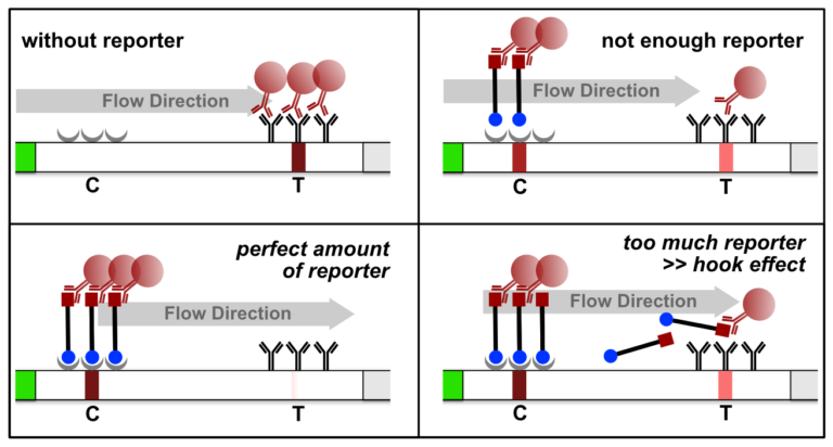
Figure 6. Reporter induced Hook Effect in the HybriDetect system.

It is necessary to understand the hook effect, to avoid misinterpretation and handling errors. According to this effect it is possible to create identical signal intensities with different concentrations of the dual labeled reporter. Furthermore, it is possible to create false negative results especially for assays, which are designed according to the described label incorporation strategy. It is crucial to limit the number of relevant labels in the LFA.

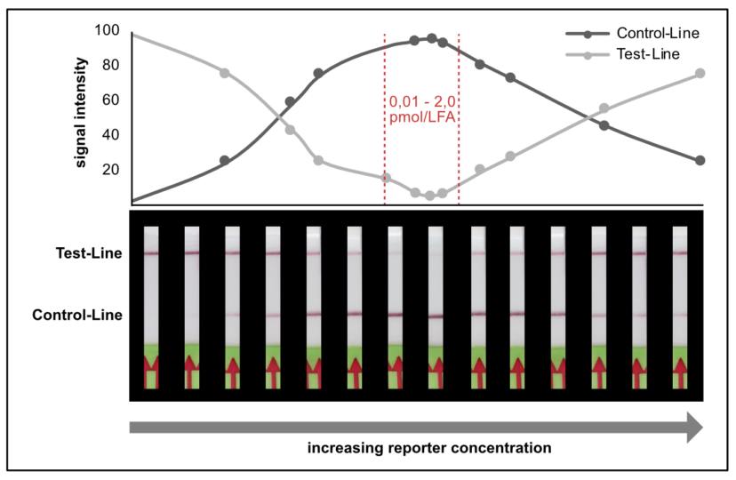
Figure 7. Idealized C- and T-line intensity depending on the reporter concentration in the LFA to illustrate the reporter induced hook effect in the HybriDetect system.

In other experimental settings we tried to determine the maximum number of labels per LFA without running into the hook effect. The following table gives a brief overview of the discussed limitations.

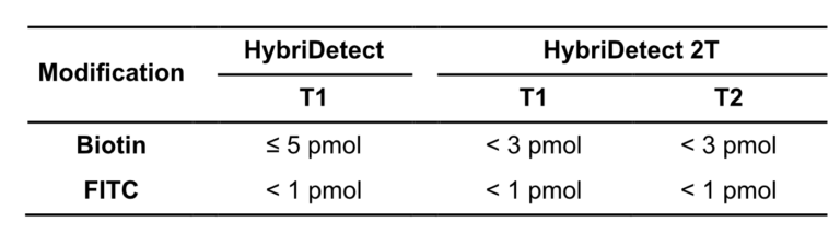
Table 2. Overview of the maximum label amounts before running into the hook effect. T-Lines are defined according to the instruction for use, and not according to CRISPR/Cas-related HybriDetect usage.

Tips and tricks for sufficient T-Line Elimination

# Good range for reporter concentration: 0,1 – 2 pmol / LFA
# Too little amount of reporter and too much reporter will result in increasing T-Line intensity >> difficult to interpret
# CAUTION >> Too much reporter induces high dose hook effect
# Long reporter sequence can result in secondary structures, or reporter-reporter interaction >> possible influence on the detectability
# Pretest new reagents and new lots

Advanced Tips

The use of 1 or 2 pmol may not be the optimum for a quick and efficient measurement of nuclease activity. If the reporter concentration in the nuclease assay deviates from T-line elimination range, the T-line intensity will be too strong for an intuitive interpretation. But there are some valuable tricks to achieve the best possible readout.

SPIKE IN: additional non cleaved reporter can be added to the Assay Buffer. Generally, you can use ssDNA reporter and increase stability of spiked assay buffer independent of the nuclease assay type. This is an easy tool for test-line intensity finetuning, if the reporter amount is not sufficient for T-line elimination. Assay buffer modification is an underrated tool in HybriDetect associated analysis.


DILUTE / less SAMPLE: at the end it’s all about pretesting your test system in the LFA context. Try to be confident using less sample or dilute before LFA. Less can be more!


SAMPLE on CRP: Try to applicate your sample directly on the Conjugate Release Pad (max. 10 µL) before you dip the stick into the assay buffer (in addition to the reporter SPIKE IN). The potentially cleaved reporter will have an advantage to bind to the mobile antibodies. Sometimes this can be beneficial to achieve sensitive test results.


TIME: Analysis time is extremely important. Two – three minutes of Lateral Flow are most frequently used in the reporter degradation dependent methods until documentation. The contemporaneous analysis start is important and can be challenging with 10 or more strips. To achieve comparable results regards analysis time, try to implement the 12-Strip-Dip-Template (can be downloaded here) in your assay procedure.

References:

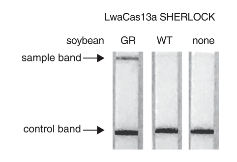
  1. Myhrvold C et al., 2018. Field-deployable viral diagnostics using CRISPR-Cas13.
  2. Abudayyeh OO et al., 2019. Nucleic Detection of Plant Genes Using CRISPR-Cas13.
  3. Sullivan et al., 2019. Rapid, CRISPR-based, Field-Deployable Detection of White Spot Syndrome Virus in Shrimp.
  4. Zhang et al., 2020. A protocol for detection of COVID-19 using CRISPR diagnostics.
  5. Metsky et al., 2020. CRISPR-based surveillance for COVID-19 using genomically-comprehensive machine learning design.
  6. Kaminsky et al., 2020. A CRISPR-based assay for the detection of opportunistic infections post-transplantation and for the monitoring of transplant rejection.
  7. Broughton et al., 2020. CRISPR–Cas12-based detection of SARS-CoV-2.
  8. Tsou et al., 2019. A CRISPR Test for Detection of Circulating Nuclei Acids.
  9. Zhang Y. et al., 2020. Evaluation of CRISPR/Cas12a-based DNA detection for fast pathogen diagnosis and GMO test in rice.
  10. Zhang Y and Ying YY, 2015. Homogeneous Immunochemical Assay on the Lateral Flow Strip for Measurement of DNase I Activity.


Author
André Breitbach
abreitbach@milenia-biotec.de
+49 (641) 948883 – 0

6 new Articles, referring to the use of CRISPR/Cas and HybriDetect

03/27
2020

6 new Articles, referring to the use of CRISPR/Cas and HybriDetect

DNA and RNA Detection with the Help of HybriDetect

New techniques have been developed for the specific detection of DNA or RNA sequences. They all have in common, that they use CRISPR/Cas12 or Cas13. The enzyme recognizes a specific target sequence. After binding to this sequence, its endonuclease activity is activated and it cleaves any reporter sequence. This technique can be easily combined with our Lateral Flow Assay HybriDetect for the visualization of the results.

1. SHERLOCK: nucleic acid detection with CRISPR nucleases
read full article

Researchers from the Broad Institute recently published an article about a CRISPR-based diagnostic platform that combines nucleic acid pre-amplification with CRISPR-Cas enzymology for specific recognition of desired DNA or RNA sequences. The described technique is named the SHERLOCK (specific high-sensitivity enzymatic reporter unlocking) method. This method can easily be combined with our HybriDetect  for fast visualization of the results.

The CRISPR/Cas-enzymes Cas12 or Cas13 are activated by binding to a specific target. When activated, the endonucleases are able to cleave nonspecific reporters.

A simple, instrument-free and portable way to readout the SHERLOCK reaction is using our LFA (lateral flow assay) HybriDetect.

The Way HybriDetect is used in the SHERLOCK Assay:

A reporter labeled with FITC on one side and Biotin on the other side has to be used. Upon activation of Cas12/Cas13 this reporter is cleaved. The biotin side of the reporter is captured by the lower band of the HybriDetect dipstick. Anti-FITC antibodies, which are labeled with gold nanoparticles, will bind on the other side of the Reporter (FITC). A dark purple line is formed at the Control band of the dipstick. Upon activation of Cas12/cas13 by binding the specific target sequence the reporter is cleaved. The gold NP-labeled antibodies, which are bound to the FITC side of the reporter will travel across the test band of the dipstick. Here they are bound by species specific antibodies and form a purple line at the 2nd position.

Reference:

Kellner M. J., Koob J. G., Gootenberg J. S., Abudayyeh O. O., Zhang F., SHERLOCK: nucleic acid detection with CRISPR nucleases. Nature Protocols (2019); V 14 (10, 2986-3012)

2. SARS-CoV-2 Detection
read full article

Due to the current COVID-19 outbreak, many labs are working on fast and easy, but still accurate ways to detect SARS-CoV-2. Since now, the EUA-approved assay developed by the US CDC is the standard test for SARS-CoV-2, it is a qRT-PCR and needs 4-6 hours.

Broughton et al. developed a CRISPR-Cas12 related assay for the detection of SARS-CoV-2 from patient RNA in 30 minutes, called DETECTR.

Upstream a reverse transcription (RT) with a Loop-mediated Isothermal Amplification (LAMP) simultaneously (RT-LAMP) has to be done. After this, Cas12 detection of specific viral sequence and cleavage of a reporter molecule is carried out. The final step is the visualization by our lateral flow test HybriDetect. Which has the main advantage of showing quick results without any instruments needed.

Clinical Testing of the DETECTR CRISPR Assay is currently ongoing.

Reference:

Broughton J. P., Deng X., Fasching C.L., Singh J., Streithorst J., Granados, A., Sotomayor-Gonzalez A., Zorn K., Gopez A., Hsu E., Gu W., Miller S., Pan C., Guevara H., Wadford D., Chen J., Chio C.Y., Rapid Detection of 2019 Novel Coronavirus SARS-CoV-2 Using a CRISPR-based DETECTR Lateral Flow Assay. medRxiv (2020); V 415

3. Detection of red tide dinoflagellate
read full article

Karenia mikimatoi is a common red tide alga, which can induce the death of fish and shellfish and therefore causes economic losses. The authors developed a new detection method using a LAMP combined with our HybriDetect for visualization. The whole procedure takes only 23 minutes from preparation of the sample to result.

Only a simple water bath or heat block is needed. The LAMP combined with HybriDetect shows is more specific and faster, compared to agarose gel electrophoresis or simple adding of SYBR green. The authors could detect the results 3 minutes after inserting the dipstick into their probe.

Reference:

Huang H. L., Gao W.F., Zhu P., Zhou C.X., Qiao L.L., Dang C.Y., Pang J.H., Yan X.J., Molecular method for rapid detection of the red tide dinoflagellate Karenia mikimotoi in the coastal region of Xiangshan Bay, China. Journal of Microbiological Methods (2020); V 168


4. Cas9-Mediated Lateral Flow Nucleic Acid Assay
read full article

Wang et al. presented a new method called CRISPR/Cas9-mediated lateral flow nucleic acid assay (CASLFA), which is one of the most convenient analytical techniques for analyzing immune response. They identified Listeria monocytogenes, GMOs and African swine fever virus (ASFV) with a detection limit of hundreds of copies of genome samples with high specificity within one hour on our HybriDetect. ASFV infected samples could be detected with an accuracy of 100% compared to RT-PCR.

The authors point out, that no laboratory environment is needed. The point-of-care-use without any technical expertise and ancillary equipment is given with this new technique.

Reference:

Wang X., Xion E., Tian T., Cheng M., Lin W., Wang H. Clustered Regularly Interspaced Short Palindromic Repeats / Cas9- Mediated Lateral Flow Nucleic Acid Assay. ACS Nano (2020), IF 13.903.


5. Detecting Plant Genes
read full article

Abudayyeh et al. presented a modified SHERLOCK detection method to quantify levels of the glyphosate resistance gene in a mixture of soybeans and to detect multiple plant genes in a single reaction. They described a field-ready SHERLOCK platform combined with Milenias HybriDetect for detection of genes in a range of agricultural applications.

Prior the Cas-reaction (20 minutes), a Recombinase Polymerase Amplification (RPA), which takes 10 minutes, is done.

The high sensitivity (single molecule, 2 aM input concentration in 1 µL sample) and specificity for single nucleotide discrimination is the major advantage of this technique. It can be used for field application, and easily combined with HybriDetect when a reporter labeled with biotin and FAM is added to the SHERLOCK reaction.

Taken together the described method is a useful platform for many biotechnological and agricultural applications.

Reference:

Abudayyeh O. O., Gootenberg J. S., Kellner M. J., Zhang F., Nucleic Acid Detection of Plant Genes Using CRISPR-Cas13. The CRISPR Journal (2019), V 2 (3, 165-171)

6. Detecting Pathogens and GMOs
read full article

Limit of rapid, low cost, user-friendly, field deployable DNA test method

Another plant application using CRISPR and HybriDetect is described by Zhang et al.. The group used a Cas12 based DNA detection method for crop disease diagnosis and a GMO test. They also used a RPA combined with Cas12 cleavage and HybriDetect for visualization.

A great advantage of this method is that it runs at 37°C and no extra instruments except filter paper and HybriDetect is needed.

The tested rice blast pathogen and Bt-rice (transgenic rice) were efficiently identified from leaf disc samples.

The described method is a rapid, low-cost, user-friendly DNA test method, which can be easily applied in field for crop disease diagnosis and GMO administration.

Reference:

Zhang Y., Zhang Y., Xie K., Evaluation of CRISPR/Cas12a-based DNA detection for fast pathogen diagnosis and GMO test in rice. Molecular Breeding (2020); V 40,11

Click here to see how to perform HybriDetect


Author
Dr. Jacqueline Hoffmann
QA — RA
jhoffmann@milenia-biotec.de
+49 (641) 948883 – 0

Lateral Flow Assays using Genome Editing Tools

04/09
2020

Diagnostic CRISPR Tools and Techniques

Lateral Flow Assays using Genome Editing Tools

What is CRISPR/Cas?

The CRISPR / Cas system acts as an immune system equivalent that is anchored in the genome of bacteria and archaea. The underlying mechanism protects the organism from phage infections by sequence specific destruction of “unknown” nucleic acids. The CRISPR/Cas-system has the ability to learn, remember and adapt. (1, 2)

This defense system is based on regions of repeating DNA-sequences, called “Clustered Regularly Interspaced Short Palindromic Repeats, also referred to as CRISPR. In addition, CRISPR-associated proteins (Cas proteins) are required for successful defense. Transcribed CRISPR-RNAs are able to guide Cas

protein(s) to the viral genome. A characteristic feature of Cas proteins is the endonuclease activity, which causes the specific degradation of viral nucleic acids. These combined features of sequence specific recognition and cutting have been used for the development of genome editing tools. (3, 4)

Figure 1. The steps of CRISPR-mediated immunity. adapted from Molecular Cell 54, April 24, 2014. (5)
Importance of Diagnostic Alternatives in the Point-of-Care Field

There is a great need for diagnostic alternatives that are suitable for simple, fast, specific, sensitive, and inexpensive early detection of pathogens. Simple handling and the avoidance of expensive and complex devices are considered to be particularly important, especially for third world countries or regions with limited lab capacities. (6, 7)

This deficiency became very clear in the years 2014 to 2016 during the Ebola outbreak (8). Today we are experiencing an even more extreme situation. The Sars-CoV-2 pandemic affects the whole world and safe, scalable diagnostics is one of the most important issues today. As part of WHO’s response to the outbreak, the R&D Blueprint has been activated to accelerate diagnostics, vaccines and therapeutics for this novel coronavirus (9).
What Makes CRISPR Associated Enzymes Attractive for Diagnostic Purposes?

In recent years, CRISPR/Cas-based detection systems have increasingly come into focus as serious diagnostic alternatives. But why are some Cas proteins particularly suitable for molecular biosensing?
Within the Cas protein classification model, all diagnostically relevant Cas proteins originate from class II. These are multidomain proteins that are guided to the target nucleic acid by associated single stranded RNAs. Another essential feature of diagnostically relevant Cas proteins is a so-called collateral activity, which occurs after a target sequence has been successfully recognized. Single-stranded RNA-/ DNA-fragments are efficiently degraded in a short period of time due to this collateral activity. And this function makes these proteins such interesting tools in diagnostics. By using molecular reporters, the collateral activity can be used for signal generation in a diagnostic approach. Reporters are short DNA- or RNA-fragments that have defined labels at their ends. Reporter cleavage leads to the generation of a detectable signal.

CRISPR/Cas-dependent Reporter Cleavage via Collateral Activity

Different Cas proteins have special characteristics and differ in some relevant criteria such as: size, recognition of nucleic acid type, collateral degradation of ssDNA or ssRNA (10). Table 1 gives a brief overview of the three diagnostically relevant Cas proteins and their specific features.

Table 1. Characteristics of commonly used CRISPR-associated Proteins for diagnostic purpose

CRISPR/Cas-Systems and Lateral Flow Readout with HybriDetect

CRISPR/Cas-based detection methods can be combined with a simple Lateral Flow Readout. Therefore the universal lateral flow platform HybriDetect is the perfect tool for a sensitive, rapid, equipment-free and simple visualization of test results (11). The general mechanism is explained in the following Figure 3.

Figure 3. General mechanism of CRISPR/Cas-mediated detection of nucleic acids via HybriDetect Lateral Flow. (A) The presence of the genetic target leads to a positive test result. (B) The absence of the specific genetic target leads to a negative test result.

The interpretation of the HybriDetect dipstick is extremely simple. If the intensity of the T-line exceeds the T-line intensity of the negative control, the test is interpreted as positive. At the same time, the C-line intensity decreases in clear positive results. The simple and intuitive interpretation of the test strips is illustrated in the following figure. About 200 copies of artificial virus RNA can be clearly detected using the CRISPR/Cas-based detection method in combination with HybriDetect lateral flow assay.

Figure 4. Detection of synthetic ZIKA virus ssRNA using SHERLOCK with 1 hour of LwaCas13a reaction, followed by Lateral Flow with Milenia HybriDetect. (12)

CRISPR/Cas-detection methods are mostly combined with pre-amplification steps. Isothermal amplification such as LAMP or RPA are the most frequently used techniques. Combinations of these methods are named SHERLOCK, DETECTR or HOLMES. Recently, it has been proved that these CRISPR/Cas-methods are able to detect pathogenic viral genomes at attomolar levels in a simple, rapid and low-equip Point-of-Care-approach using the Milenia HybriDetect.

# Detection of Zika Virus , SHERLOCK_CRISPR/Cas13-System (12,13)
# Detection of Dengue Virus, SHERLOCK_CRISPR/Cas13-System (12)
# Detection of Human Papillomavirus (HPV) -16 and -18, CRISPR/Cas12a-System (14)
# Detection of SARS-CoV-2, CRISPR/Cas12a-System, CRISPR/Cas13-System (15,16)

References

Jinek M, Chylinski K, Fo nfara I, Hauer M, Doudna JA, Charpentier E. A programmable dual‐RNA–guided DNA endonuclease in adaptive bacterial immunity. Science. 2012;337(6096): 816‐821. DOI: 10.1126/science.1225829, https://www.ncbi.nlm.nih.gov/pmc/articles/PMC6286148/pdf/nihms-995853.pdf

Wright AV, Nuñez JK, Doudna JA. Biology and applications of CRISPR systems: harnessing nature’s toolbox for genome engineering. Cell. 2016;164(1‐2):29‐44. DOI: https://doi.org/10.1016/j.cell.2015.12.035

Singh V, Gohil N, Ramírez García R, Braddick D, Fofié CK. Recent advances in CRISPR‐Cas9 genome editing technology for biological and biomedical investigations. J Cell Biochem. 2018;119(1):81‐94. https://doi.org/10.1002/jcb.26165

Zetsche B, Gootenberg JS, Abudayyeh OO, et al. Cpf1 is a single RNA‐guided endonuclease of a class 2 CRISPR‐Cas system. Cell. 2015;163(3):759‐771. Doi: 10.1016/j.cell.2015.09.038.https://www.ncbi.nlm.nih.gov/pmc/articles/PMC4638220/pdf/nihms725840.pdf

Barrangou, R. and Marraffini, L. CRISPR-Cas Systems: Prokaryotes Upgrade to Adaptive Immunity (2014). Molecular Cell 54, 234-244. Original Image: http://sitn.hms.harvard.edu/flash/2014/crispr-a-game-changing-genetic-engineering-technique/

Zumla A, Al-Tawfiq J, Enne V, Kidd M, Drosten C, Breuer J, Muller M, Hui D, Maeurer M, Bates M, Mwaba P, Al-Hakeem R, Gray G, Gautret P, Al-Rabeeah A, Memish Z, Gant V. Rapid point of care diagnostic tests for viral and bacterial respiratory tract infections-needs, advances, and future prospects. The Lancet Infectious Diseases. 2014 vol: 14 (11) pp: 1123-1135. Doi: 10.1016/S1473-3099(14)70827-8.https://www.ncbi.nlm.nih.gov/pmc/articles/PMC7106435/pdf/main.pdf

Chen H, Liu K, Li Z, Wang P. Point of care testing for infectious diseases.Clinica Chimica Acta. 2019 vol: 493 pp: 138-147. Doi: 10.1016/j.cca.2019.03.008. https://doi.org/10.1016/j.cca.2019.03.008

Pollock NR, Wonderly B. Evaluating novel diagnostics in an outbreak setting: lessons learned from Ebola. J Clin Microbiol. 2017;55(5):1255‐1261. https://jcm.asm.org/content/jcm/55/5/1255.full.pdf

WHO, A research and development Blueprint for action to prevent epidemics. Source: https://www.who.int/blueprint/en/

Aman R, Mahas A, Mahfouz M. Nucleic Acid Detection Using CRISPR/Cas Bio-sensing Technologies. ACS Synthetic Biology. 2020. doi:10.1021/acssynbio.9b00507. https://pubs.acs.org/doi/10.1021/acssynbio.9b00507

James A, Todd S, Pollak N, Marsh G, Macdonald J.Ebolavirus diagnosis made simple, comparable and faster than molecular detection methods: Preparing for the future.Virology Journal. 2018 vol: 15 (1). https://doi.org/10.1186/s12985-018-0985-8.

Gootenberg J, Abudayyeh O, Kellner M, Joung J, Collins J, Zhang F. Multiplexed and portable nucleic acid detection platform with Cas13, Cas12a, and Csm6. Science vol. 360, pp: 439-444 (2018). doi: 10.1126/science.aaq0179. https://www.ncbi.nlm.nih.gov/pmc/articles/PMC5961727/pdf/nihms965860.pdf

Myhrvold C, Freije C, Gootenberg J, Abudayyeh O, Metsky H, Durbin A, Kellner M, Tan A, Paul L, Parham L, Garcia K, Barnes K, Chak B, Mondini A, Nogueira M, Isern S, Michael S, Lorenzana I, Yozwiak N, Macinnis B, Bosch I, Gehrke L, Zhang F, Sabeti P. Field-deployable viral diagnostics using CRISPR-Cas13. Science vol. 360, pp: 444-448 (2018). Doi: 10.1126/science.aas8836. https://science.sciencemag.org/content/360/6387/444/tab-pdf

Tsou J, Leng Q, Jiang F. A CRISPR Test for Detection of Circulating Nuclei Acids. Translational Oncology. 2019 vol: 12 (12) pp: 1566-1573. doi: 10.1016/j.tranon.2019.08.011 https://www.sciencedirect.com/science/article/pii/S1936523319304140?via%3Dihub

Broughton J, Deng X, Yu G, Fasching C, Streithorst J, Granados A, Sotomayor-Gonzalez A, Gopez A, Hsu E, Gu W, Miller S, Pan C, Wadford D, Chen J, Chiu C, Chiu C.Rapid Detection of 2019 Novel Coronavirus SARS-CoV-2 Using a CRISPR-based DETECTR Lateral Flow Assay. PREPRINT server: bioRxiv. Doi: 10.1101/2020.03.06.20032334. https://www.medrxiv.org/content/10.1101/2020.03.06.20032334v2

Metsky H, Freije C, Kosoko-Thoroddsen T, Sabeti P, Myhrvold C. CRISPR-based surveillance for COVID-19 using genomically-comprehensive machine learning design. PREPRINT server: bioRxiv. 2020 pp: 2020.02.26.967026. doi: 10.1101/2020.02.26.967026. https://www.biorxiv.org/content/10.1101/2020.02.26.967026v2.full.pdf

Author
André Breitbach
abreitbach@milenia-biotec.de
+49 (641) 948883 – 0

Lateral Flow Test Development for COVID-19 based on Nucleic Acid Detection

10/26
2020

Lateral Flow Test Development for COVID-19 based on Nucleic Acid Detection

Amplicon detection via the lateral flow test Milenia “HybriDetect”

Recently Milenia Biotec GmbH received several customer inquiries related to COVID-19 detection, due to the fact, that HybriDetect may be used for the detection of RPA, LAMP or PCR amplicons. Following up these inquiries, we did a literature search on the SHERLOCK (Specific High Sensitivity Enzymatic Reporter UnLOCKing) technique, which is a powerful tool to detect genetic information coming from viruses using CRISPR.

What is HybriDetect?

The HybriDetect is a lateral flow dipstick (LFD), which is capable to detect different molecules, including gene amplification products, proteins and antibodies. A commonly used application for our test is the detection of 


gene amplification products resulting from PCR, LAMP or RPA. The underlying genetic information can be detected regardless which kind of organism need to be detected. Finally, labeled primers must be introduced in the amplification step, so that the resulting fragments are labeled and can be detected by the HybriDetect dipstick.

Results can be reported within 5 minutes after lines become visible on the test strip.
If you want to learn more about HybriDetect, for example information on the limit of detection, see our other blog articles.

COVID-19 Detection Methods

At the moment, the standard COVID 19 test is a qRT-PCR (quantitative real time polymerase chain reaction). Obviously, there are several reasons why qRT-PCR test cannot be used as a POCT (point of care test). First of all, it takes several hours to do a qPCR test, second you need highly trained personnel and third special equipment is essential. In the current situation, with the entire world dealing with the COVID-19 outbreak, it is of great interest to develop rapid tests for POC testing of the new corona virus to help people in sparsely populated areas where laboratories are difficult to reach.

Since the outbreak of COVID-19 at the end of 2019 several tests for SARS-CoV-2 detection have been developed and approved. But most of them show several disadvantages:

# Expensive laboratory equipment is needed
# High level technical expertise is required
# Access to reagents is difficult
# Expensive

With the currently available rapid Antigen tests, a result is available after 15 minutes outside the lab. But the Antigen tests are detecting viral proteins, and are therefore less sensitive as a test on DNA level.

Due to these reasons, several groups are working on alternative tests for the quick, easy and low cost detection of SARS-CoV-2 with very little equipment needed.

Why do we need more rapid tests?

The head of the WHO Tedros Adhanom Ghebreyesus already told on March 16th, that there has not been an urgent enough escalation in testing. His key message on this day was: test, test, test.

The most important point is to detect local outbreaks as soon as possible and control the spread of the COVID-19 virus through quarantine measures. The effect of the high test capacities could be seen in South Korea. They rely on early PCR tests to record lots of cases, even mild and symptom free ones. Tests are offered in drive troughs and walk troughs. Consequently, the number of infections in South Korea stayed relatively low.

At a school in Mecklenburg-West Pomerania, in Germany all teachers and students, even their parents are offered free tests to identify and contain infections early. The tests are voluntary and offered twice a week. Mecklenburg-West Pomerania is planning to do the COVID-19 tests in every school, to get back to “normal life conditions” quickly.

These two examples show the high need for frequent testing. Obviously, obtaining fast test results is one of the biggest advantages of point-of-care compatible rapid tests. Generating quick test results without the need of sending the samples to an external laboratory allows quick reaction and can help to control local outbreaks.

Here we give a short overview of the latest publications dealing with the rapid detection of

COVID-19 combined with the HybriDetect Lateral Flow Method for visualization.

COVID 19 testing and CRISPR

Promising testing methods, which are based on isothermal amplification in combination with a CRISPR-mediated detection (SHERLOCK (1), DETECTR (2)) have been developed.

SHERLOCK

The group of the inventor of the SHERLOCK method Feng Zhang and collegues, based in the United States, described a method for COVID-19 detection using CRISPR. The scientists were able to detect synthetic COVID-19 virus RNA fragments between 20 and 200 aM (10-100 copies per µl of input). They used purified RNA as Input for an RT-RPA before the Cas13 assay. The whole method takes less than one hour.

The following picture shows an example of the COVID-19 detection using our HybriDetect.

Fig. 1: Covid-19 testing. Detection of the Covid-19 S gene with HybriDetect Strips. First four dipsticks show positive result.

For more information see the original paper:

https://www.broadinstitute.org/files/publications/special/COVID-19%20detection%20(updated).pdf

Zhang et al give a precise description, how the SHERLOCK technique is done in nature protocol:

http://dx.doi.org/10.1038/s41596-019-0210-2

STOPCovid – improved SHERLOCK method

Due to the fact, that the method described above needs two reaction steps (isothermal amplification and CRISPR-reaction) it is still complex and cross-contamination in an uncontrolled environment can happen quickly. For these reasons, Zhang and colleagues recently published an update (preprint, not peer reviewed) on the SHERLOCK method, which can be performed in one step (3) and is a promising rapid COVID 19 detection method.

The SHERLOCK method was continously improved in the last few years to make specific POC tests possible:

2016: Detection of Cas13 by Zhang lab
2018: Improvement of the Cas enzymes Cas12b and Cas13 (SHERLOCKv2)
2019: First paper on the specific detection of DNA and RNA through CRISPR-Cas from clinical relevant samples. Step-by-step instructions with RPA
2020: STOPCovid: One Pot reaction combines LAMP and SHERLOCK to detect Sars-CoV-2

The so-called STOP (SHERLOCK Testing in One Pot) method is an improvement of the previous SHERLOCK application. This test allows a turnaround time of an hour from sampling to the report of the resuls. The very simple handling underlines the potential as a point-of-care test (POCT) for COVID 19 testing.

General information about STOP:

~Results in 40 to 70 minutes
~Detection of 100 copies of viral genome
~Saliva or nasopharyngeal swabs as input
~Specific detection
~Single temperature, one fluid handling step, visual readout
~Nearly no equipment

Fig. 2 Equipment needed for STOP. Beside a simple heat block, some tubes, pipettes and tips, HybriDetect lateral flow strips need to be used for COVID19 testing.

One pot reaction in three simple steps

Step 1: 10 mins at 22°C or at 60°C → lysis of virus-containing patient sample (add Proteinase K inhibitor) or 5 mins at 95°C

Step 2: 1 hr at 60°C → detection of viral RNA using STOPCovid reaction

Step 3: 2 mins at 22°C → visual read out of the detection results by eye using HybriDetect

Fig. 3: STOPCovid in five steps – from sample to result.

If you want to learn more about the lateral flow readout of HybriDetect in combination with CRISPR, read our related article: Lateral Flow Readout for CRISPR/Cas-based detection strategies

Combining RT-LAMP, Cas12b and HybriDetect

The authors used a RT-LAMP (loop-mediated isothermal amplification) for RNA-transcription followed by simple isothermal DNA-amplification, which works at 55-65°C. For this reason they needed a thermostable Cas-Protein, which is active at 60°C. They found Cas12b from Alicyclobacillus acidiphilus (Aap) to be stable at this temperature. Out of 29 primer sets for detecting SARS-CoV-2 the scientists found LAMP-compatible primer sets. The next step was to find the right guide RNA (gRNA), which was figured out comparing 18 gRNAs.

During another optimization it was discovered that the addition of taurin resulted in an improvement of the reaction kinetics. After RT-LAMP and CRISPR-reaction (Step 3 in Figure 2) HybriDetect can be dipped directly into the reaction tube at room temperature and visual read out can be done after 2 minutes.

The Authors provide all the information needed to do the STOPCovid test in their paper: LAMP Primers, gRNA sequence, Cas12b-Protein Sequence, etc.

Validation of STOPCovid

The method was validated on 17 patient samples. It could detect 12 positive and 5 negative samples, 2 of 3 replicates were scored positive in infected patients. STOPCovid was able to detect all positive and all negative samples (Fig. 3) compared to the gold standard method RT-qPCR.

Fig. 4: Validation of STOPCovid. Results for 17 unique SARS-CoV-2 nasopharyngeal swab samples. Quantification of the band intensity ratio of lateral flow results.

STOPCovid.v2

The inventors of STOPCovid are constantly improving the Covid-19 detection method for rapid testing. In theire latest update on the method they described a magnetic bead purification method. With this approach, they could reduce the sample extraction time to 15 minutes. (Fig. 5)

The researchers compared the STOPCovid.v2 with the gold standard RT-qPCR. The result was a reduction of viral RNA that was 600 times of the input needed for the RT-qPCR. The STOPCovid.v2 has a limit of detection (LOD) similar to a cycle-theshold (Ct) value of 40.3. StopCovid.v2 was tested on 202 SARS-CoV-2 positive and 200 SARS-CoV-2 negative samples. The test result showed a sensitivity of 93.1% and a specificity of 98.5%. The positive samples were detected in 15 to 45 minutes.

Read the original article here: https://www.nejm.org/doi/full/10.1056/NEJMc2026172?query=featured_home
What is the clue about the updated version of SHERLOCK?

Taken together the striking advantage of this method is its simplicity. The 3 steps, shown above are very easy to do, even for untrained personnel. The authors even showed that saliva samples worked as input for the reaction. This makes the method performable for lay users. Another advantage is the low-tech equipment approach, which has a major impact on the POC-compatibility.

Scientists who are interested in testing the protocol can get more information and test kits at STOPCovid.science.

The protocol is not authorized by the FDA (food and drug administration), it is not for clinical purposes.

Read the original article here: https://www.medrxiv.org/content/10.1101/2020.05.04.20091231v1.full.pdf

DETECTR

Broughton et al. already published a paper, how to detect SARS_CoV-2 using LAMP, Cas12 and our HybriDetect within 30 minutes. They are using isothermal pre-amplification with primers published by the WHO and CDC. As the authors mentioned in the paper, this application would be a helpful tool for POCT testing in emergency departments, airports etc..

Here you can find a video from the McGovern Institute describing the SHERLOCK technique in combination with the HybriDetect in General

In summary, rapid testing is being expanded to stop the spread of Covid 19. Scientists in the United States and India (read more about the Feluda Test) developed new tests on Genome level, which are nearly as fast as rapid antigen tests, but more sensitive. With these new approaches, departments of health could be relieved and the public health improved.

References

  1. Zhang et al., 2020. A protocol for detection of COVID-19 using CRISPR diagnostics.
  2. Broughton et al., 2020. CRISPR–Cas12-based detection of SARS-CoV-2.
  3. Joung et al., 2020. Point-of-care testing for COVID-19 using SHERLOCK diagnostics

Author
Dr. Jacqueline Hoffmann
QA — RA
jhoffmann@milenia-biotec.de
+49 (641) 948883 – 0

HybriDetect and Temperature Effects

03/20
2020

HybriDetect and Temperature Effects

Using higher Temperatures: Part of the NAFLIA Toolbox

Higher Temperatures: Stability aspects and assay Parameters

The general robustness is a characteristic feature of lateral flow devices (LFD’s). Even during longer transports and extreme weather conditions LFD’s remain stable. Temperature is an important factor when it comes to robustness. Therefore our HybriDetect test strips are regularly tested at higher temperatures.

Especially in the development of NALFIA’s (Nucleic Acid Lateral Flow Immunoassays), the temperature can influence the test result significantly. Higher temperatures can also be understood as tool in the assay procedure to achieve certain results. The hybridization of amplificates is classified as particularly interesting. The use of a defined temperature is a crucial parameter to maintain specific hybridizing conditions.

Higher Temperatures: Performance Test

For this reason, Milenia Biotec GmbH took a closer look at this issue: The universal test strips HybriDetect (MGHD1) and HybriDetect 2T (MGHD2 1) were examined under different conditions with dilution series of labelled dsDNA. The purified labelled amplificates were placed into running buffer and warmed to the desired temperature. Test strips were prewarmed as well for 30 minutes. After rewarming Lateral Flow Analysis was initiated. After five minutes signals were documented. The overall results are shown in the following figure.

Figure: Influence of higher temperatures to LFA-performance of HybriDetect test strips. Red triangles indicate temperature related nonspecific signals on test lines.

The results illustrate the robustness of the dipsticks. Even at 65 °C, a sensitive analysis is possible without a visible loss of performance. Nonspecific signal appear over 70°C, especially on the testline 2 of the HybriDetect 2T (red triangles in the figure). This effect is easy to explain. Like most LFD’s the HybriDetect platform includes antibodies. If these antibodies are exposed to temperatures over 70°C, denaturation takes place. This results in non-specific signals.

However, the full functionality of the HybridDetect test strips was demonstrated in the tested range of 20°C to 65 °C. Temperature should be considered as an additional tool in assay development. Try to profit from this feature!

Author
André Breitbach
abreitbach@milenia-biotec.de
+49 (641) 948883 – 0

Detecting Viruses – Major Pathogens in Aquaculture

08/12
2019

Detecting Viruses

Major Pathogens in Aquaculture

Several investigative techniques have been developed to detect viruses causing severe fish and shrimp diseases. The methods used are LAMP and RPA combined with our Lateral Flow Dipstick (LFD) for DNA Detection: HybriDetect. The addressed viruses cause diseases with a high mortality rate and therefore have a huge economic impact in fish farming industries (1-6).

Short Method Descriptions
LAMP – Loop-mediated isothermal amplification

LAMP is an isothermal DNA amplification method and thus can be done without using a thermocycler. LAMP based amplification procedures are usually done in the temperature range between 60°C and 60,001°C.

In general, two sets of primers (two outer and two inner primers) are used to identify six regions on the target gene (high specificity compared to PCR). LAMP uses Bst DNA polymerase large fragment, which has a high strand displacement activity. Compared to PCR, LAMP is very rapid (1 h).

RPA – Recombinase Polymerase Amplification

RPA is as specific as PCR amplification but much faster and can be done at temperatures between 37 and 42°C in just 10 minutes. RPA uses a recombinase, a single-stranded DNA-binding protein (SSB) and a polymerase. The recombinase pairs the primers to the homologue target DNA sequences. SSB stabilizes the resulting D-loop. DNA synthesis is initiated by the DNA polymerase.

LFD – Lateral Flow Dipstick – HybriDetect

The HybriDetect is a lateral flow dipstick (LFD), which is able to detect different molecules, such as gene amplification products, proteins and antibodies. A commonly used application for our test is to detect gene amplification products resulting from PCR, LAMP or RPA. Therefore, labeled primers must be used during the amplification step, so that the resulting DNA fragments are labeled and can be detected by the HybriDetect dipstick. Results can be reported within 5 minutes after lines become visible on the test Strip. HybriDetect is working with aqueous solution and does not contain toxic reagents such as EthBr.

Detecting Viruses – Applications
Infectious spleen and kidney necrosis virus (ISKNV) detection
read full article

Ding et. al. developes a LAMP combined with the Milenia HybdriDetect 2T Lateral Flow Dipstick (LFD) for the detection of ISKNV (Infectious spleen and kidney necrosis virus), one of the most important pathogens in aquaculture, especially in China. The ISKNV causes high mortality in many freshwater and marine fish. The authors developed a test with a detection limit of 10 copies for the large cytoplasmic dsDNA virus. The method is rapid, sensitive, simple and has a high economic impact. The sensitivity was 1 000 fold higher than in comparable methods , like LAMP-AGE (4).

Cyprinid herpes virus 2 (CyHC-2) detection
read full article

CyHV-2 causes Herpesviral heamoatopoietic necrosis (HVHN) in carp aquaculture and is responsible for huge economic losses in China, USA and Australia. There is no effective prevention and the disease causes mortality up to 100 %. It is crucial, that HVHN is detected in a very early stage of the disease. Wang et al developed a RPA combined with our HybriDetect which can be done in just 15 minutes at 38 °C. The method is 100 times more sensitive than others and the authors couldn’t detect any cross reactions with other aquatic viruses (3).

Cybrinid herpes virus 3 (CyHC-3) and Koi Herpes Virus (KHV) detection
read full article

A LAMP-LFD (HybriDetect) method for the specific detection of cybrinid herpes virus 3 (CyHC-3) and Koi Herpes Virus (KHV) was invented by Soliman and El-Matbouli. This method can detect amounts of 10 fg DNA (30 copies vg) within one hour. It is much faster than the commonly used PCR (3 hours) and 10 to 100 fold more sensitive (5).

Detection and Differentiation of Carp oedema virus (CEV) and koi herpes virus (KHV)
read full article

With the current tests on the market, it is often difficult to determine between CEV and KHV, which both are common carp viruses. In 2017 a rapid and accurate tool was invented by Soliman and Matbouli to detect and differentiate CEV and KHV. The authors used RPA combined with HybriDetect (LFD) in their new assay which is very fast (60 min) compared to current methods (10 and 7 hours). The test can be used in field situation to reduce spread of the viruses (2).

Spring Viremia of Carp Virus (SVCV) Detection
read full article

SVCV is a cyprinid pathogenic virus, which usually needs to be detected in a lab. Most virus outbreaks can be seen in fishery banks. The invented detection method (LAMP with HybriDetect) is suitable for field-detection in aquaculture and can detect up to 860 fg DNA. The authors claim, that a rapid and accurate diagnosis of the virus is vital to prevent the spread of the virus and to minimize economic losses. Many commonly used techniques are time consuming or show cross reactions with other viruses; therefore a simple, but still accurate method like LAMP-LFD is needed (1).

Shrimp Taura Syndome Virus detection
read full article

Kiatpathomchai et al also used a combination of LAMP with LFD to detect the Shrimp RNA Virus Taura Syndrome Virus (TSV). This virus has a big economic impact regarding shrimp farming. The method developed by the authors is very quick (total assay about 70 min.). The sensitivity is comparable to other commonly used methods for RT-PCR detection of TSV (6).


Advantages

In summary the combination of LAMP or RPA combined with HybriDetect (LFD) Shows a number of advantages:

  • * Equivalent or even better sensitivity compared to commonly used PCR methods
    * Much faster
    * No cross reactions to other aquatic viruses
    * Field-application
    * Nearly no equipment/machines needed
    * Cost effective


Literature

  1. Comparison of Three Terminal Detection Methods Based on Loop Mediated Isothermal Amplification (LAMP) Assay for Spring Viremia of Carp Virus (SVCV). (2019). Turkish Journal of Fisheries and Aquatic Sciences, 19(9), 805–816Soliman, H., & El-Matbouli, M. (2018)
  2. Rapid detection and differentiation of carp oedema virus and cyprinid herpes virus-3 in koi and common carp. Journal of Fish Diseases, 41(5), 761–772. https://doi.org/10.1111/jfd.12774
  3. Wang, H., Sun, M., Xu, D., Podok, P., Xie, J., Jiang, Y., & Lu, L. (2018). Rapid visual detection of cyprinid herpesvirus 2 by recombinase polymerase amplification combined with a lateral flow dipstick. Journal of Fish Diseases, 41(8), 1201–1206. https://doi.org/10.1111/jfd.12808
  4. Ding, W. C., Chen, J., Shi, Y. H., Lu, X. J., & Li, M. Y. (2010). Rapid and sensitive detection of infectious spleen and kidney necrosis virus by loop-mediated isothermal amplification combined with a lateral flow dipstick. Archives of Virology, 155(3), 385–389. https://doi.org/10.1007/s00705-010-0593-4
  5. Soliman, H., & El-Matbouli, M. (2010). Loop mediated isothermal amplification combined with nucleic acid lateral flow strip for diagnosis of cyprinid herpes virus-3. Molecular and Cellular Probes, 24(1), 38–43. https://doi.org/10.1016/j.mcp.2009.09.002
  6. Kiatpathomchai, W., Jaroenram, W., Arunrut, N., Jitrapakdee, S., & Flegel, T. W. (2008). Shrimp Taura syndrome virus detection by reverse transcription loop-mediated isothermal amplification combined with a lateral flow dipstick. Journal of Virological Methods, 153(2), 214–217. https://doi.org/10.1016/j.jviromet.2008.06.025

Author
Dr. Jacqueline Hoffmann
QA — RA
jhoffmann@milenia-biotec.de
+49 (641) 948883 – 0

Hygiene monitoring in breweries

03/11
2019

Hygiene monitoring in breweries

Results in just 2 hours – Immediate corrective actions possible!

The quality of beer is controlled in breweries at each individual level of the production process. The presence of beer spoilage microorganisms is associated with an elevated risk to beer quality. Microbial contaminations create a negative impact on the taste, the texture and the smell of the final product. Contamination can occur over the entire production process, although the area of the filling machine is the main spot for contamination. For this reason, filling machines are cleaned intensively on a regular basis and the success of the cleaning process is controlled via close hygiene monitoring.

During this process, swabs are taken from different places on the illing machine, especially in the area of the fillers, and are transferred to a selective culture medium in order to monitor the potential bacterial growth. In the case of slow-growing or hard-to-cultivate bacteria being present in the swab, it can take days, up to

weeks, until a positive result from a culture can be reported. In this event, initiation of causal cleaning activities cannot be initiated.

For this reason, we are presenting a method that allows detection of beer spoilage bacteria directly from swabs within 2 hours! In this setting, the detection of obligate anaerobic bacteria of the genus Megasphaera and Pectinatus are of special interest. The basis of this method is the combination of a rapid extraction of the bacteria from the swabs, followed by PCR amplification. More information about the procedure is available here.

If you have questions related to the detection of beer spoilage microorganisms from swabs, or you would like to receive a demonstration in your brewery, please fill in the Contact Form and send it to us.

Author
Dr. Ralf Dostatni
Managing Director of Milenia Biotec GmbH
rdostatni@milenia-biotec.de
+49 (641) 948883 – 0

SARS-CoV-2 RBD Proteins (501Y.V1/V2/V3) for COVID Variants of UK, South Africa, and Brazil

SARS-CoV-2 RBD Proteins (501Y.V1/V2/V3) for COVID Variants ofUK, South Africa, and Brazil

Product Cat.No. Varient Mutation Source
SARS-CoV-2 (COVID-19) S RBD (N501Y) Protein 11-064 UK 501Y.V1 N501Y Mammalian
SARS-CoV-2 (COVID-19) S RBD (K417N, E48K, N501Y) Protein 11-065 South Africa 501Y.V2 K417N, E484K, N501Y Mammalian Cells
SARS-CoV-2 (COVID-19) S RBD (E484K, K417T, N501Y) Protein 11-066 Brazil 501Y.V3 E484K, K417T, N501Y Mammalian Cells
SARS-CoV-2 (COVID-19) S RBD (E484K) Protein 11-067 501Y.V2/V3 E484K Mammalian
SARS-CoV-2 (COVID-19) Spike (D614G) Trimer Protein 92-748 501Y.V2 D614G Human Cells
SARS-CoV-2 (COVID-19) Spike (D614G) S1 Protein 92-746 501Y.V2 D614G Human Cells

B.1.1.7 Lineage of SARS-CoV-2 (2019-nCoV)

In September of 2020 a new lineage of SARS-CoV-2, known as B.1.1.7, was discovered in the United Kingdom. This lineage was found to have developed 14 lineage-specific amino acid replacements and 3 deletions prior to its discovery. It appears that the B.1.1.7 is now evolving at a rate similar to other SARS-CoV-2 lineages which have a rate of mutation of about one to two mutations per month (Duchene et al. 2020).

One of the mutations associated with this lineage is a N501Y in the spike protein of the virus. It is believed that this mutation is able to increase the spike protein’s affinity for the host ACE2 receptor (Starr et al. 2020) and it has been associated with increased infectivity and virulence (Gu et al. 2020). B.1.1.7 viruses have also been shown to have a P681H mutation in the cleavage site of spike protein. This location is one of the residues that make up the furin cleavage site between S1 and S2 in spike. The S1/S2 furin cleavage site has been shown in animal models to promote viral entry into respiratory epithelial cells and transmission (Hoffmann et al. 2020; Peacock et al. 2020; Zhu et al. 2020). The spike proteins of this lineage has also been shown to have a deletion at amino acids 69-70. This mutation in the receptor binding domain of spike is a recurrent deletion that has been found in various lineages associated with SARS-CoV-2 (McCarthy et al. 2020; Kemp et al. 2020). Outside of spike, a Q27 stop mutation truncates the ORF8 protein of the virus, rendering the protein inactive. An ORF8 deletion at amino acid 382 has a mild effect on virus replication in human airway cells (Gamage et al. 2020). The B.1.1.7. lineage also has five synonymous mutations in ORF1ab and one synonymous mutation in the M gene.

ProSci Inc. has developed specific antibodies with the peptide immunogen including the mutation site and these antibodies can be used for Western Blot, ELISA, IHC/IF, and other immunoassays.

[Edited from Rambaut et al, 2020]

Cat.No. Antibodies
9087 SARS-CoV-2 (COVID-19) Spike RBD Antibody
9091 SARS-CoV-2 (COVID-19) Spike S1 Antibody (cleavage)
9095 SARS-CoV-2 (COVID-19) Spike S1 Antibody (cleavage)

SARS-CoV-2 Spike Recombinant Proteins (D614G)

Product Cat.No. Soure Fusion Tag Sequence
SARS-CoV-2 (COVID-19) S-Trimer Protein Recombinant Protein (D614G) 92-748 Human Cells C-6 His Tag Cys15 – Gln1208 (D614G)
SARS-CoV-2 (COVID-19) S1 Protein Recombinant Protein (D614G) 92-746 Human Cells C-10 His Tag Gln14 – Arg685 (D614G)

SARS-CoV-2 (COVID-19) S RBD Mutant Recombinant Proteins

Product Cat.No. Soure Fusion Tag Sequence
SARS-CoV-2 (COVID-19) Spike RBD Recombinant Protein 92-750 Human Cells C-6His Tag Arg319 – Phe541 (F342L)
SARS-CoV-2 (COVID-19) Spike RBD Recombinant Protein 92-751 Human Cells C-6His Tag Arg319 – Phe541 (N354D)
SARS-CoV-2 (COVID-19) Spike RBD Recombinant Protein 92-752 Human Cells C-6His Tag Arg319 – Phe541 (V367F)
SARS-CoV-2 (COVID-19) Spike RBD Recombinant Protein 92-753 Human Cells C-6His Tag Arg319 – Phe541 (R408I)
SARS-CoV-2 (COVID-19) Spike RBD Recombinant Protein 92-754 Human Cells C-6His Tag Arg319 – Phe541 (A435S)
SARS-CoV-2 (COVID-19) Spike RBD Recombinant Protein 92-755 Human Cells C-6His Tag Arg319 – Phe541 (K458R)
SARS-CoV-2 (COVID-19) Spike RBD Recombinant Protein 92-756 Human Cells C-6His Tag Arg319 – Phe541 (G476S)
SARS-CoV-2 (COVID-19) Spike RBD Recombinant Protein 92-757 Human Cells C-6His Tag Arg319 – Phe541 (V483A)
SARS-CoV-2 (COVID-19) Spike RBD Recombinant Protein 92-758 Human Cells C-6His Tag Arg319 – Phe541 (D364Y)
SARS-CoV-2 (COVID-19) Spike RBD Recombinant Protein 92-759 Human Cells C-6His Tag Arg319 – Phe541 (V341I)
SARS-CoV-2 (COVID-19) S RBD-SD1 Recombinant Protein (V367F) 92-742 Human Cells C-6 His Tag Arg319 – Ser591 (V367F)
SARS-CoV-2 (COVID-19) Spike RBD-SD1 Recombinant Protein (N354D, D364Y) 92-743 Human Cells C-6His Tag Arg319 – Ser591 (N354D, D364Y)
SARS-CoV-2 (COVID-19) Spike RBD-SD1 Recombinant Protein (W436R) 92-744 Human Cells C-6His Tag Arg319 – Ser591 (W436R)

SARS-CoV-2 (COVID-19, 2019-nCoV) Research

References

Duchene, Sebastian, Leo Featherstone, Melina Haritopoulou-Sinanidou, Andrew Rambaut, Philippe Lemey, and Guy Baele. 2020. “Temporal Signal and the Phylodynamic Threshold of SARS-CoV-2.” Virus Evolution 6 (2): veaa061.

Gamage, Akshamal M., Kai Sen Tan, Wharton O. Y. Chan, Jing Liu, Chee Wah Tan, Yew Kwang Ong, Mark Thong, et al. 2020. “Infection of Human Nasal Epithelial Cells with SARS-CoV-2 and a 382-Nt Deletion Isolate Lacking ORF8 Reveals Similar Viral Kinetics and Host Transcriptional Profiles.” PLoS Pathogens 16 (12): e1009130.

Gu, Hongjing, Qi Chen, Guan Yang, Lei He, Hang Fan, Yong-Qiang Deng, Yanxiao Wang, et al. 2020. “Adaptation of SARS-CoV-2 in BALB/c Mice for Testing Vaccine Efficacy.” Science 369 (6511): 1603–7.

Hoffmann, Markus, Hannah Kleine-Weber, and Stefan Pöhlmann. 2020. “A Multibasic Cleavage Site in the Spike Protein of SARS-CoV-2 Is Essential for Infection of Human Lung Cells.” Molecular Cell 78 (4): 779–84.e5.

Kemp, S. A., D. A. Collier, R. Datir, S. Gayed, A. Jahun, M. Hosmillo, Iatm Ferreira, et al. 2020. “Neutralising Antibodies Drive Spike Mediated SARS-CoV-2 Evasion.” Infectious Diseases (except HIV/AIDS). medRxiv. https://doi.org/10.1101/2020.12.05.20241927.

McCarthy, Kevin R., Linda J. Rennick, Sham Nambulli, Lindsey R. Robinson-McCarthy, William G. Bain, Ghady Haidar, and W. Paul Duprex. 2020. “Natural Deletions in the SARS-CoV-2 Spike Glycoprotein Drive Antibody Escape.” Microbiology. bioRxiv.

Peacock, Thomas P., Daniel H. Goldhill, Jie Zhou, Laury Baillon, Rebecca Frise, Olivia C. Swann, Ruthiran Kugathasan, et al. 2020. “The Furin Cleavage Site of SARS-CoV-2 Spike Protein Is a Key Determinant for Transmission due to Enhanced Replication in Airway Cells.” Cold Spring Harbor Laboratory. https://doi.org/10.1101/2020.09.30.318311.

Starr, Tyler N., Allison J. Greaney, Sarah K. Hilton, Daniel Ellis, Katharine H. D. Crawford, Adam S. Dingens, Mary Jane Navarro, et al. 2020. “Deep Mutational Scanning of SARS-CoV-2 Receptor Binding Domain Reveals Constraints on Folding and ACE2 Binding.” Cell 182 (5): 1295–1310.e20.

Zhu, Yunkai, Fei Feng, Gaowei Hu, Yuyan Wang, Yin Yu, Yuanfei Zhu, Wei Xu, et al. 2020. “The S1/S2 Boundary of SARS-CoV-2 Spike Protein Modulates Cell Entry Pathways and Transmission.” Cold Spring Harbor Laboratory. https://doi.org/10.1101/2020.08.25.266775.

Lyophilised or liquid?

Lyophilised or liquid?

Lyophilised – no expertise needed

RPA has a number of advantages over PCR. The isothermal technique dispenses with thermocycling instruments and offers rapid results within five to 20 minutes, making it ideally suited to point-of-care testing and experiments away from the laboratory. Freeze-drying the reagents into a lyophilised pellet format enables us to put RPA into the hands of non-experts; buffers and magnesium acetate are simply added to the pellet with the template mixture and the reaction starts. Rehydration is the key step, and applying a little heat to the reaction will benefit sensitivity. Results can even be obtained using body heat, and in West Africa – where access to heating technology is limited – an ambient temperature of 30 °C is sufficient to carry out plant pathogen testing using our technology. Lyophilising the material into dry pellets increases the stability at these temperatures, reducing the need for cold storage and encouraging use outside the lab, from veterinary diagnostics on the farm to mobile testing for infectious diseases.

“Liquid RPA kits can be used in both high and low volume experiments, allowing reactions to take place in miniaturised formats – nanolitres to microlitres – which remain challenging to PCR.”

Liquid – high throughput and low volumes

High throughput, lab-based applications in industry and academia are at the other end of the spectrum, and a lyophilised format would, in many cases, be entirely inappropriate. We therefore also provide RPA reagents in a wet, glycerol-stabilised format, which simplifies the production process by cutting out the freeze-drying step.However, the liquid versions of the kits need to be stored at a low temperature to keep them stable, something that would not be suitable for point-of-care testing in the field. Offering RPA in a liquid format also opens the door to applications that would simply not be feasible using a lyophilised pellet. Liquid RPA kits can be used in both high and low volume experiments, allowing reactions to take place in miniaturised formats – nanolitres to microlitres – which remain challenging to PCR. This format is equally advantageous for larger volume reactions from 500 µl to 1 ml. It takes a comparatively large amount of energy and time to heat 1 ml of liquid during PCR, whereas RPA has less intensive requirements, operating at a constant, lower temperature.

Two formats, multiple applications

Both the lyophilised and liquid formats function in the same manner, and we are excited to be witnessing the adoption of the technology across a broad range of sectors, from microfluidics to water hygiene to biodefence. There are dozens and dozens of assays that researchers have created and published in peer-reviewed journals; so many that I can no longer keep up with reading all the papers as I used to. Whatever your application, our customer service team is always on hand to talk through any questions and discuss the most suitable RPA format for your application. Please get in touch and we’ll be happy to help. 

From Biochemistry to Brewing

From Biochemistry to Brewing

Name: Chris Walowski
Qualifications: BS Biochemistry and MS Biochemistry, 10+ Years Brewing Experience
Occupation: Head Brewer at Trustworthy Brewing Co.
Hobbies: Beer, Fly Fishing, Fishing, Gardening, Fermenting Vegetables ie. pickles, peppers, etc. Cooking

“Brewing beer is an art and a science,” says Chris Walowski of Trustworthy Brewing Co. “The art is making a product that people enjoy and the science is making that product taste the same exact way every time.” With his background in biochemistry and over ten years of experience in the brewing industry, Chris knows what he’s talking about. From big dreams and studying biochemistry at CSULB, to crafting cult-favorites as the Head Brewer of Trustworthy Brewing Co., Chris shares his unique experience how he became the brewmaster he is today.

How did you get into brewing beer and when did you decide to fully pursue it as a career?

During college, I had a part-time job at Trader Joes and was introduced to new styles and flavors of beer than just the Bud, Miller or Coors. I began to start exploring new beer styles and quickly learned that finding craft beer was very difficult. I saw a show on TV about home brewing and there happened to be a home-brew store very near to my parent’s house. The TV show made it seem very easy since there was a store so close by, it was convenient and since it was really hard finding fresh craft beer, I decided to take matters into my own hands. I began brewing at home during my undergraduate degree at CSULB. It slowly turned into a fun hobby and then to a passion. I started brewing multiple times a week and winning awards at local competitions. I then started grad school and my accolades drew some attention. I was approached by a startup brewery in LA to help out and brew some beer. I, of course, said yes and quickly transitioned to head brewer and was working part-time. I finished my MS and was offered a full-time salaried position at a new brewery. I took the job and began my brewing career.

“Being able to understand the fermentation pathways and how biochemical pathways produce off flavors allows me to troubleshoot issues much more efficiently.”

How has your background in biochemistry given you an edge in your career? Can you give a few examples?

Having a background in science and biochemistry has given me a very definitive edge in my career. “Brewing” beer is a bit of a misnomer in my opinion. The yeast makes the beer! I am merely a steward. My job is to make sure the yeast is happy and comfortable and that fermentation is done correctly. Sure, I am the one who crafts the recipe for the beer, but without a heathy and proper fermentation, the beer will be undrinkable. Being able to understand the fermentation pathways and how biochemical pathways produce off flavors allows me to troubleshoot issues much more efficiently. I am comfortable around a microscope and other scientific instruments. All the ingredients we use are spec’d for all sorts of parameters like protein content, enzyme content, alpha acids, esters and phenols. Having my degree allows me to quickly analyze and understand what I’m working with.

Is brewing beer an art or a science?

It is both! What I like to say is the art of making beer is making a killer recipe and flavor profile that a consumer will enjoy. The science of making beer is using your knowledge to achieve that goal and also being able to make the same consistent product batch over batch while your ingredients are changing.

“…the art of making beer is making a killer recipe and flavor profile that a consumer will enjoy.”

What role does fermentation play in the brewing process?

Fermentation is the most important process. Well besides sanitation… Fermentation is the process that makes the beer. Before fermentation, the beer isn’t that good tasting but after fermentation, it is a different story.

From a scientific standpoint, what is the difference between ‘good’ and ‘bad’ beer?

Most of the flavor flaws are directly caused by particular organic molecules that get produced through metabolic pathways during fermentation. Acetaldehyde and diacetyl, for example, are produced by yeast under stressed conditions. Some particular minerals weren’t in high enough concentrations, a low amount of viable cells were added or not properly monitoring fermentation temperatures can all lead to off flavors.

What makes beer bitter?

Beer is bitter by “alpha acid” compounds found in hops. When the beer is boiled, you are actually conducting a reflux. The precursor molecules found in the hops isomerize during the boiling process and cause it to become bitter.

What is the ‘correct’ way to pour a beer? What goes on at a chemical level?

I am not a stickler when it comes to how a beer is poured. I mostly prefer it to be cold, carbonated and poured in a clean glass. I do like to see a bit of head on the poured beer and I want the foam to remain on top of the beer while I drink it. On a chemical level, beer protein foam stability is a huge subject of scientific research. Scientists know mostly what proteins are involved but exactly how they interact is a still a mystery. Also, foam and foam proteins interaction only form once. So the more foam you generate, during production, packaging and serving, the less amount will actually stay on top of the beer.

What are two fun science facts about beer that not many people know?

1.Enzymes were discovered by a scientist who was researching beer. The scientist lysed a yeast cell and discovered that there was something inside the yeast that continued to ferment the beer. He called these “things” En-Zymes which translates to In-Yeast from Latin. 2. Louis Pasteur was the scientist who discovered that yeast was the organism responsible for fermenting beer. Before Pasteur, it was scientific dogma that the sugar in beer spontaneously decomposed to alcohol and CO2. It almost ruined his career trying to convince the scientific community and this fact was overshadowed by his other contributions to science and food stability.

What are some common inaccurate myths about beer?

The color of the beer indicates strength and that low alcohol beer has less flavor. Consumers often assume that Guinness is heavier and has more calories but Guinness is around 3.5% ABV which is lower in ABV than Bud and it has less calories. This example disproves both myths.

What is one of the biggest lessons you learned as you left your research position and launched a brewing company?

The biggest lesson was being able to balance what I wanted to brew or what I personally find interesting with what will sell or what the customer wants. I always want to brew interesting styles or with odd ingredients and sometimes the beer just doesn’t sell. Which can be disappointing, but I try to sneak in some more interesting styles like Lagers, sours and dark beers in my onsite taproom to help educate consumers and introduce them to different styles of beer.

“I like making people happy and I like to put a smile on someone’s face because they enjoyed a beer I made.”

What is the most rewarding part of what you do?

The most rewarding part of my job is interacting with customers. I like making people happy and I like to put a smile on someone’s face because they enjoyed a beer I made.

Any advice for someone wanting to start brewing their own beer?

Do your homework! Develop a business plan and be able to roll with the punches. Also always remember that you are not in the business of making beer, you are in the business of SELLING beer. There is a big difference and selling beer isn’t for everyone.

What’s your favorite beer?

I typically enjoy most hoppy styles of beer, like an IPA or hoppy lager. I like beer that is drinkable, I want to be able to drink a whole pint without it feeling like a chore.

Yeast’s Key Role

As much as yeast is essential to brewing and fermentation, it also plays a key role in Zymo Research’s history.