How Yale Monitors COVID-19 Surges in Connecticut’s Wastewater

How Yale Monitors COVID-19 Surges in Connecticut’s Wastewater

Predicting Outbreaks Before They Happen

Pandemics present a unique civic challenge for public health officials. How do you monitor infection levels in large groups of people who are very close together in a repeatable way, using limited labor and reagents?

The answer lies beneath our feet. Sewers carry the viral components shed by infected individuals to wastewater treatment plants, where those components settle with solid waste into an unsavory but informative component: sludge.

While the purification of nucleic acids from sludge is a known but difficult process, the logistics of doing so regularly and accurately at scale have few precedents. To find the best way forward, Annabelle Pan, Jordan Peccia, and Alessandro Zulli of Yale’s Environmental Engineering Program began a CDC-funded project. The goal? To actively monitor SARS-CoV-2 RNA levels from sludge samples taken from wastewater treatment facilities across the state of Connecticut.

In theory this would allow the team to detect community outbreaks as they began, giving public health officials the forewarning to take proper action. “Testing municipal wastewater provides similar information to randomly and anonymously testing thousands of people, but it’s much cheaper,” explained Annabelle Pan. “For many places around the world, this may make wastewater monitoring a more realistic and affordable option than comprehensive testing programs.”

“Testing municipal wastewater provides similar information to randomly and anonymously testing thousands of people, but it’s much cheaper.”

-Annabelle Pan

Research Technician, Yale School of Engineering & Applied Sciences

As expected, the initial push was a race to tie together a dozen loose ends. “The first month or so of this project was spent gathering samples, figuring out protocols, and ironing out all kinks.” said Alessandro Zulli. When everything finally came together, the results were anything but uncertain: “The first time we aggregated the data for New Haven and saw a curve that fit the cases to that point back in April was mind blowing.”

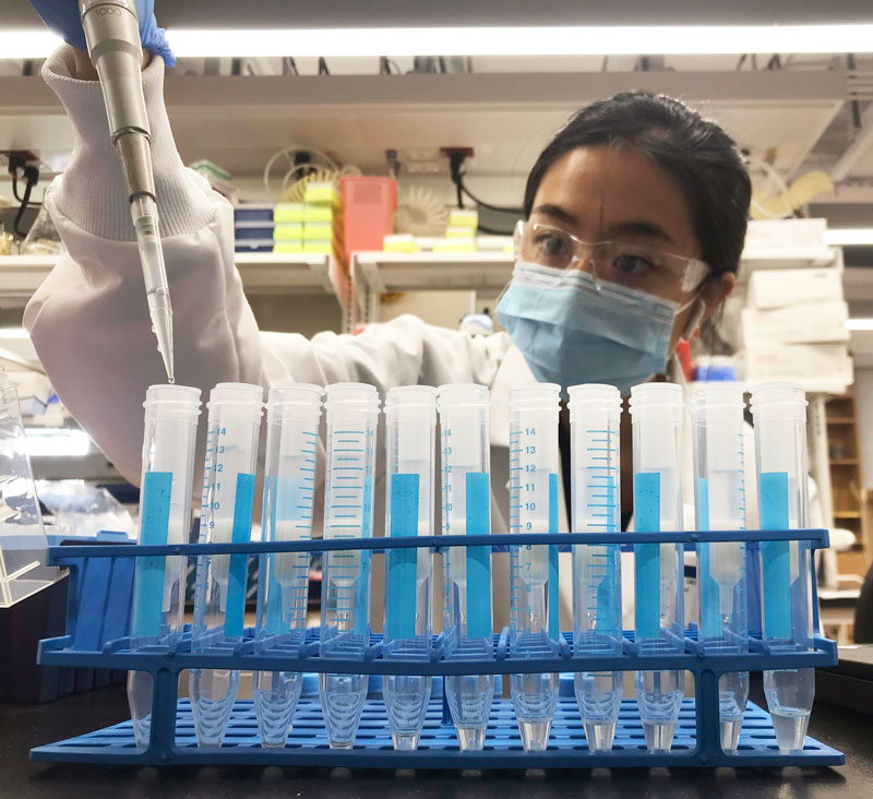
Annabelle Pan, Yale Environmental Engineering Program


With now-validated methods, the team had to scale the initiative to the state level. Though the science was sound, the logistics involved were complex. “Sludge testing isn’t super fancy science, but putting together all the pieces to make it work requires some coordination,” explained Pan. “You need people at a wastewater treatment plant who are willing to take samples for you, people who can transport those samples to a lab in a matter of hours, and people running extractions and PCR.”

“One daily primary sludge sample from the New Haven, CT wastewater treatment plant represents a population of 200,000 people.”

-Annabelle Pan

Research Technician, Yale School of Engineering & Applied Sciences

Each sample received was able to provide insight into high volumes of people: “One daily primary sludge sample from the New Haven, CT Wastewater Treatment Plant represents a population of 200,000 people,” said Pan. These results were made publicly available using a web reporting tool, so health officials and private individuals alike could use it to guide their actions.

But the tangle of local logistics was not the only challenge the team faced when trying to obtain and maintain these live results. Like many scientists, they were impacted by the supply chain shortages caused by the very pandemic they were trying to monitor. “We experienced a supply chain issue in October when the kit we originally used went out of stock indefinitely,” said Pan. “That was quite stressful because we had to scramble to find a replacement kit.”

Eventually the team found the Quick-RNA Fecal/Soil Microbe Microprep Kit suitable for their needs, “Getting a nice ~30,000ng of RNA out of the first extraction we did using the Zymo kit was truly uplifting and relieving,” said Pan. The kit enabled the team to process more samples at once, allowing the testing to be implemented with minimal resources while maximizing coverage. While making their process significantly more time efficient had a new benefit, as it allowed them to more easily comply with COVID era protocols and social distancing.

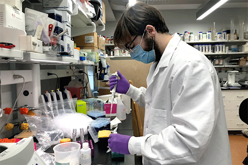
Alessandro Zulli, Yale Environmental Engineering Program

With their extraction kit and workflow validated, the team was able to begin state-wide monitoring. Helping control this global pandemic is a stressful task for any researcher, but the Yale team found gratitude in their role during difficult times. “It’s given me some purpose throughout this pandemic, and allowed me to contribute to efforts to stop the spread,” said Pan. “I’m glad for the chance to apply previously learned skills to a project this important.”

The availability of the data has also helped locals assess the best course of action, “My friends used this data to weigh being more cautious back in late October, when New Haven’s wastewater started to show a spike in covid RNA,” shared Pan.

Local public health officials have likewise used this data to take strategic action, meeting with the team twice a week. When RNA levels rose to concerning levels, the Mayor of Stamford implemented a robocall to advise residents on how to best avoid the virus.

This near-live aspect of the data is one of its most useful attributes for individuals and policymakers alike. “The value of this project has really been in providing real-time data that isn’t subject to the lags inherent to testing for cases, and for confirming/predicting trends in cases,” said Alessandro.


“The value of this project has really been in providing real-time data that isn’t subject to the lags inherent to testing for cases.”

-Alessandro Zulli

PhD Student, Yale Environmental Engineering Program

Of course, in the midst of this success the team has not forgotten that ever-present followup to successful science: repeatability. Would other universities be able to offer these services to the municipalities in which they reside? The team thinks so.

“I would say that most large institutions have the resources necessary to implement a program like this already,” said Alessandro. “It’s an incredible way to not only benefit their students, but the larger population surrounding the university.”

This project used the Quick-RNA Fecal/Soil Microbe Microprep Kit to extract viral RNA from sludge. Zymo Research has since upgraded and optimized viral RNA extraction from wastewater; the new Zymo Environ Water RNA Kit increases RNA yields from wastewater by 8-fold with its unique Water Concentrating Buffer. Zymo Research proudly provides a variety of solutions for environmental monitoring specially designed for organizations monitoring COVID-19 surges in wastewater.

Learn More About The Zymo Environ Water RNA Kit:

Current Bottlenecks With COVID-19 Testing

Current Bottlenecks With COVID-19 Testing

Four months into 2020, COVID-19 has changed the world. Currently, the US and Europe are the epicenters of the disease with cases and deaths rising exponentially while many countries have imposed strict lockdown measures in order to try to control the spread of the disease.

Through these trends, the only country that has been successful at “flattening the curve” is South Korea. They have managed to control the outbreak without instituting the strict lockdowns that have been seen in nearly all countries around the globe.

The South Korean success story can be attributed to one strategy in particular: testing.

South Korea was able to institute a massive testing campaign that identified and isolated those who were positive. They created drive-thru testing locations, customized phone booth testing locations, deployed biotechnology companies to scale up reagent manufacturing, and tracked mobile phone data to determine the past whereabouts of COVID-19 positive patients.

All of this was critical since it can take up to 14 days for someone who has the virus to show symptoms. Due to this long incubation period, testing is crucial in controlling the spread of COVID-19. Widespread testing allows us to identify and isolate those who are positive and prevent them from spreading the virus.

“The testing they did in South Korea was very important in controlling their outbreak,” said Dr. Angela Caliendo, an infectious diseases professor at Brown University’s Alpert Medical School. These strategies were so successful in curbing the spread of COVID-19 that as of March 2020, over 100 countries have asked South Korea to assist with their testing programs.

This system of rampant testing resulted in 4099 tests being administered per million people in South Korea by March 9th.  By comparison, the US has lagged behind in testing quite considerably. Even though the two countries had their first case of COVID-19 at very similar dates – January 15th for the US and January 20th for South Korea – the US had only administered 26 tests per million people by March 9th. The effects of this can be seen now in the total number of COVID-19 cases. South Korea has had 196 total cases per million people while the United States has 741 total cases per million people.

Effective, widespread testing is crucial to controlling the spread of COVID-19. However, there have been many issues and bottlenecks in getting the scale of testing to where it needs to be. The current system in the United States is dysfunctional and is not allowing the development of an adequate response to the COVID-19 crisis. Below, each step in the typical COVID-19 testing workflow is detailed along with the obstacles that need to be resolved in order to expand testing and save more lives from this disease.

Sample Collection

The first step in testing is to collect a sample. A patient has to go to a hospital, clinic, or testing center/drive-thru and meet with a healthcare professional who performs the sampling. With a sterile swab, the healthcare worker takes a nasopharyngeal (inside the nose) or oropharyngeal (back of the throat) sample. In swabbing these areas, the goal is to collect any biological material that has recently been within the lungs (where COVID-19 replicates). Once collected, the swab is stored and transported/shipped at 2-8 °C or at -70 °C  on dry ice to a clinical testing lab.

At first step in the testing workflow, there are several issues that need to be resolved in order to streamline the process.

Shortage of Swabs
The biggest problem is that the United States doesn’t have enough swabs available to collect samples from everyone who needs a test. The shortage of swabs is due to the increased demand around the globe, as swabs are needed for most COVID-19 testing.

Swabs, in particular, have an exacerbating circumstance. Copan, one of the largest manufacturers of swabs, is located in Lombardy, Italy, which was hit very hard by the virus which limited the global supply of swabs even further. Although Copan has been allowed by the Italian government to continue production amid the countrywide lockdown, they will still need to generate millions of swabs in order to meet the demand for COVID-19 testing in the United States.

The other problem with swabs is that they have to be used properly in order to yield accurate results. The correct technique requires the swab be inserted deep within the nose or throat and rotating the swab several times (which is not a comfortable experience for the patient). If the swab does not pick up enough of the virus, it will lead to a false negative result. Studies from China have shown that the false negative rate can be as high as 30%.

While nasopharyngeal swabs are the most commonly used method to collect patient samples, other sample sources can be processed just as easily. The FDA states that the COVID-19 diagnostic panel can be tested on upper and lower respiratory specimen samples which includes sputum. Sputum, or phlegm, is the mucousy substance that is secreted by cells deep within the respiratory tract. Since sputum samples are produced by the patient from deeper within the respiratory tract, it is more likely to pull up the virus from where it resides compared to swabs. The swabs can only reach the back of the throat or nasal cavity whereas sputum is expelled from deeper within the respiratory tract, giving a higher chance for the viral particles to be collected.

Recent studies have supported this by showing that sputum and fecal samples were able to return more positive COVID-19 results than swabs. These findings indicate that rate of false negatives may decrease with the adoption of sputum sampling.

In order to expand and improve the testing system, there needs to be more methods of sample collection. Currently, swabs are the only devices that have been approved for use by the FDA, but approving new collection devices for use could solve stock issues with sample collection devices and could potentially lead to more accurate test results.

Higher Risk of Infection

Current sample collection methods have healthcare workers collect a sample directly from the patient. This requires close contact with the patient and puts the worker at a greater risk of infection. This issue is only compounded by the shortage of PPE at many hospitals and testing locations.

This particular portion of the COVID-19 testing workflow is slowly changing with many hospitals and testing sites taking actions to

protect their workers from the disease. Firstly, the FDA has announced that self-administered testing, under the supervision of a healthcare worker, is allowed at testing locations for symptomatic patients. This helps protect healthcare professionals from having direct contact with the patient. A study by United Health Group found that of 500 patients who self-administered swab collection tests, COVID-19 was detected in more than 90% of positive patients, which was consistent with the clinically administered test.

Other solutions have included drive-through sample collection sites which allow potentially infected patients to stay isolated in their cars, away from other patients and workers. This eliminates the need for patients to enter the hospital for COVID-19 testing and risk infection by sitting in a crowded waiting room of symptomatic patients to deliver their sample.


Healthcare workers collect COVID-19 samples at a drive-through testing site in Florida.

The last solution that could further protect clinicians and patients would be to keep the potentially infected people away from the healthcare facilities by performing sample collection at home. This would entail the collection device being mailed to the patient who then self-administers the test and then mails it back to a lab for processing. Currently, the FDA has not authorized any at-home testing or sample collection for COVID-19, but has been sent proposals from several companies.

One of the biggest reasons for the slow approval has been concern that patients are not able to self-collect samples correctly. While instructions on how to collect samples may help to reduce confusion for the general public, the FDA needs further proof that patients are able to collect samples at home without increasing the risk for false-negatives.

Another is reason for the delays in approval lie with the stability of the sample during shipment. The COVID-19 test detects the RNA from the virus, and because RNA tends to be much less stable than DNA, there is a concern that the RNA can degrade. If this happens, the RT-qPCR would not be able to amplify the viral RNA and the test would be negative.

Because of this, it is crucial that the appropriate stabilization reagents are used in potential at-home collection workflows. They can prolong the stability of the RNA for up to 30 days at ambient temperatures meaning that samples can be effectively collected and transported without worry of a false negative result. By stabilizing the RNA, these reagents are able to detect very small amounts of the virus allowing more sensitive assays to be performed.

As we continuously optimize the testing workflow, these considerations need to be taken into account in order to effectively protect both clinicians and patients.

RNA Extraction for COVID-19 Testing

Once the sample arrives in the lab, the RNA needs to be purified from the sample with an extraction kit manually or with an automated liquid handler.

Shortage of RNA Extraction Kits
Just like swabs, there is a dramatic shortage of the reagents required to purify and analyze the viral samples for COVID-19 testing. Once the patient sample arrives in the lab, the RNA needs to be extracted. This process requires a series of chemical buffers that lyse, bind, and purify the viral RNA. Some labs make their own reagents to extract RNA, but most use commercial kits that take hours off the processing time.

With the increasing demand for COVID-19 testing, many labs are struggling to find reagents from their suppliers that are required to process the test. This then delays test results and increases the risk of further spread of the virus as patients may assume the delayed result means they are negative for COVID-19.

The situation has gotten so bad that some scientists have taken to twitter to request donations from other scientific labs. Since the RNA extraction kits are used in a wide range of applications, there were several labs, companies, and research institutes that were not analyzing COVID-19 that could help offset the critically low level of extraction kits.

As one of the largest manufacturers of RNA extraction kits, Zymo Research has increased their manufacturing capabilities to help support the massive need for additional reagents. They will be producing enough of their kits to enable millions of coronavirus tests each month.

COVID-19 Detection
The purified samples contain the host RNA and any COVID-19 RNA (if present). To detect the viral RNA in the sample, a molecular biology technique called real-time reverse transcription polymerase chain reaction(rRT-PCR). This system amplifies the small amount of viral COVID-19 RNA present to thousands of copies, so that may be detected. This  makes this assay extremely sensitive.

If there is no virus present within the sample, there will be nothing for the rRT-PCR to amplify and the machine will show a negative result.  

Shortage of Reagents
Like with all other supplies in the testing workflow, the detection reagents for the rRT-PCR are also in short supply. There are shortages of the detection buffers, dyes, and enzymes will that dramatically slowdown the time to get results to patients.

Scaling Up

Typically, when a lab begins to process a high number of samples they turn to automated solutions and move away from processing samples by hand. This is done by robotic platforms that greatly increase the throughput of a lab, allowing hundreds or even thousands of samples to be processed in a single day.

Stalled Implementation of Automation

As the testing demand for COVID-19 has grown, it has strained processing labs, and many of these laboratories are looking toward high-throughput automated workflows to keep up with the flood of patient samples that need to be processed. However, these automated workflows usually require lengthy amounts of time before a lab can fully utilize the machine because it has to be set up appropriately.

First, the machine needs to be delivered and installed. Since these are large and sensitive pieces of equipment, this process can take up to a week. Once the robot has been set up, every movement of the robot has to be coded in (or scripted) so that it can manipulate the samples appropriately and isolate the viral RNA. Just like coding an app on your phone, the scripting process is very detailed and time-consuming. Every movement of the robot has to be detailed and entered into the machine: the volume of reagent added at each step, where the reagent is dispensed, how many times the sample should be mixed, and more. Once these commands are correctly entered into the liquid handler, the script has to be tested and then optimized as needed.

From here, the script needs to be validated with test samples to ensure the device can function properly. Once it has been validated, the lab can finally begin processing patient samples on the machine.

This process is time-consuming and arduous for many labs and can take several weeks to complete from the time the device is purchased to the first patient samples being processed on it. In the COVID-19 era, these delays simply hold back labs from testing as many samples as they need to.

The best solution to avoid these delays in implementing automation is to use an automated liquid handler that comes preloaded with scripts that have already been configured and optimized for viral RNA extraction, like the DreamPrep NAP. This device delivers a load-and-go system with a user-friendly interface that dramatically decreases the amount of time needed to begin processing samples. With this device, labs can quickly and easily scale up their operations to accommodate testing for a much higher number of COVID-19 samples.


In the world of biology, this workflow is not very complicated. Theoretically, this workflow could function seamlessly with samples flowing in from patients and being processed by the lab with results delivered in 1-2 days. However, the global pandemic has placed a severe strain on the entire testing workflow. Just as with hospitals being overburdened and reaching capacity, testing labs around the world are feeling this same effect. Because of this, there is a severe delay in the implementation and expansion of critical testing services, which adds days to the processing time.

This strain on the testing system is unlike anything the field has ever seen. The industry needs rapid implementation of new solutions to combat the virus from all angles.

With scientists around the world working feverishly to expand production, implement new testing workflows, and streamline processes, the United States is slowly ramping up the amount of tests that can be administered. However, none of these tactics will be effective without the help of the public. Just as the public can flatten the curve for hospitals by staying home, that same effect applies to testing laboratories. With most of the public staying home, they are already helping to resolve the testing bottleneck by decreasing the number of samples that need to be processed. As the world comes together to fight the virus, every single person has a part to play.

See related blogs:

How To Increase Plasmid Yield

How To Increase Plasmid Yield

Sometimes, plasmid purification just doesn’t go according to plan. Whether a vector is kept at low copy number, low culture density, or culture overgrowth, this guide will help you navigate your purification woes and determine the best way to boost your plasmid yields from E. coli cultures.

Increase the Amount of Culture Processed

Sometimes the simplest way for how to increase plasmid yields is to just input more raw material. While this is simple enough in theory, there are a few considerations to observe before you start adding more culture to your plasmid preps. Most plasmid prep kits have limitations on the amount of culture they can process, and these limitations will vary based on the copy number of your plasmid as well.

The efficacy of column-based plasmid purification doesn’t just depend on how much plasmid is loaded onto the column, but also the total amount of biomass being loaded as well. If you want to scale up your plasmid purification, try using a kit designed for high inputs of culture, such the ZymoPURE II Midipreps and Maxipreps, which can process more culture.

Increasing the amount of culture processed leads to higher total yield of plasmid DNA if kit parameters are adjusted accordingly to accommodate higher biomass.

Optimize Your Bacteria

Sometimes particular E. coli strains are sub-optimal for plasmid extraction. If you are experiencing low yields for your plasmid prep, double check that the strain you’re using is best for plasmid propagation. Some strains are more optimized for protein expression than for efficient DNA replication. Others have unwanted byproducts, such as carbohydrates or endonucleases, which can co-purify with plasmids. When possible, stick to tried and tested strains like E. coli DH5α which contain mutations to lack certain endonucleases and increase plasmid stability. Thus, these modified E. coli strains are used as workhorses for molecular cloning and plasmid production.

Use Optimal Growth Conditions

Never inoculate culture straight from your bacterial stock. Always start with a single colony that is then grown as a starter culture. This ensures that your culture is derived from the same genotype and is not a mix of different colonies which may not have the same characteristics.

Also, be sure to double check your growth conditions. While many guides will recommend 12-16 hour cultures, every E. coli strain is slightly different in its growth rate and final density. Some require different temperatures, longer incubation times, faster shaking speeds, or more oxygenation.

For example, the maximum culture volume should not exceed 1/5 the total volume of the growth flask or alternatively, growth in baffled flasks can be used to increase aeration, and thus, culture growth. Also, take note of the shaking speed of the culture, with 200-250 rpm typical.

Additionally, total plasmid yield can vary depending on the type of culture broth used, typically Luria-Bertani broth (LB) or Terrific Broth (TB). Don’t be afraid to experiment with your growth conditions to see which gives you the best plasmid yield.

Purification from cultures grown in highly enriched media such as Terrific Broth (TB) yield higher amounts of plasmid DNA per ml of culture than standard Luria-Bertani Broth (LB).

Optimize Selective Pressure and Yield

There are a couple ways to ensure high yield plasmid preparations from E. coli cultures by using antibiotics for selective pressure.

The first is the antibiotic selection required for any molecular cloning experiment. It’s imperative to ensure the correct amount of antibiotic is present in your culture, otherwise the lack of selection pressure will cause your culture to start to dilute out the plasmid during cell division.

The second method uses chloramphenicol, an antibiotic that halts protein synthesis and decouples it from plasmid replication, when culturing strains containing a plasmid with a relaxed origin of replication. Chloramphenicol treatment can stop protein production but allow the E. coli  to continue to “amplify” the plasmids, resulting in increased yields during plasmid purification1.

One method for plasmid amplification uses an inhibitory amount of chloramphenicol (170 µg/ml) added to a culture, which is then incubated further until plasmid purification (typically the next day)2. A variation of this method that reports higher plasmid yield uses lower amounts of chloramphenicol (10-20 µg/ml) added to exponentially growing cells that are subsequently incubated overnight prior to plasmid purification3. Alternatively, another study demonstrated increased plasmid yield by growth in the presence of sub-inhibitory concentrations of chloramphenicol (3-5 µg/ml) from the time of culture inoculation until plasmid was harvested the next day4. Note that these treatments only work for chloramphenicol-sensitive cells and plasmids that do not encode for chloramphenicol resistance.

Bringing It Full Circle

Next time you’re stumped about your plasmid purification, apply these tips on how to increase your plasmid yields and optimize your experiment. Whether it be increasing the volume, changing your growth conditions, adding more selective pressure, or using the best isolation technologies, there is always optimization to be done to increase your plasmid yields!

Learn how to collect high purity plasmid DNA:

References:

1. Ausubel, FM, et al. Current Protocols in Molecular Biology, (2003)
2. Sambrook, J, Fritsch EF, Maniatis T, Molecular Cloning: a laboratory manual, 2nd edition. Cold Springs Harbour Laboratory Press, Cold Springs Harbour, New York, 1989
3. Frenkel L, Bremer H, Increased Amplification of Plasmids pBR322 and PBR327 by Low Concentrations of Chloramphenicol, DNA 5, num 6, 539-544 (1986)
4. Begbie S, et al., The Effects of Sub-Inhibitory Levels of Chloramphenicol on pBR322 Plasmid Copy Number in Escherichia coli DH5a Cells, Journal of Experimental Microbiology and Immunology 7, 82-88 (2005)
5. https://www.zymoresearch.com/


What Are Transformation, Transfection & Transduction?

What Are Transformation, Transfection & Transduction?

One of the pillars of modern day molecular biology uses techniques to manipulate DNA sequences (such as plasmids, knockout gene constructs, etc.) and introduce them into a host cell to test their effects. However, getting the DNA into cells can take different routes. Those unfamiliar with the field may be wondering “what is plasmid transduction?” Or have heard the terms transformation, transfection, and transduction, but are uncertain as to the differences and similarities between these techniques. Although these terms have some overlap, and so their usage is often confusing or incorrect.

What Is Plasmid Transformation?

Transformation is, simply put, the process of altering a cell’s genetic code through the uptake of foreign DNA from the environment. Plasmid transformation is used to describe the (non-viral) horizontal gene transfer of plasmids between bacteria. While transformation likely happens in the natural world, scientists have harnessed this process to their own ends, enabling replication of lab-manipulated plasmids and expression of desired recombinant DNA sequences.

The process is relatively simple; scientists make the membranes of bacterial cells permeable to DNA either through chemical means or via electrical stimulation. These cells, now termed ‘competent cells,’ will readily uptake plasmid DNA from their surroundings. Once the DNA molecule of interest is introduced to these competent cells, the bacteria have now been plasmid transformed. The transformed cells then can be selected from the untransformed cells by inclusion of an antibiotic to kill off the untransformed cells. Typically, this occurs as the plasmid will express an antibiotic resistance gene to protect the transformed cells and ensure maintenance of the plasmid over time and cell divisions. In the process, many replicons of the plasmid will be created and passed to daughter cells.

What Is Plasmid Transfection?

Transfection is a type of plasmid transformation, typically that of animal cells, instead of bacteria. This process is a bit more complicated than your run-of-the-mill transformation, as many lab-cultured eukaryotic cells do not natively uptake and replicate foreign DNA. Still, scientists have discovered many ways in which plasmids and other foreign DNA can be introduced to cells.

Much like methods for bacteria, there are both chemical and physical methods of transfection produce transient holes in the cell membrane and get uptake of foreign DNA. These methods work similarly to the those outlined for bacterial transformation, as they all are designed to make the cell membrane more permeable. The method by which they do so is different from bacteria, though, instead using cationic lipids, micelles, lasers, or even particle guns. These methods have their pros and cons, but ultimately will depend on the resources available and the preference of the researcher.

What Is Transduction?

The final prominent method, transduction, is unique from the other two methods. Transduction is the process of using a virus to mediate the delivery of DNA fragments or plasmids into a cell, either prokaryotic or eukaryotic. This technique harnesses the natural function of viruses to inject DNA into the infected host, but with a twist. Scientists can modify the viral nucleic acids to contain specific DNA sequences of interest. There are many different types of viruses that can be manipulated to introduce recombinant nucleic acids into host cells. For example, bacteriophage introduce DNA into bacteria, and lentiviruses or adenoviruses into human cells. Using these modified viruses, researchers incorporate foreign DNA into the host genome (such as using lentiviruses or bacteriophage) or transiently express desired recombinant nucleic acids (such as using adenoviruses).

In order to perform a transduction, you need a cell-line of interest and a virus that infects that cell line. This method can be more difficult than the other methods discussed here, since the virus must be grown and maintained in culture, sometimes needs to be modified to be non-infectious to humans, and the DNA of interest must be packaged into the viral particle before infection of new host cells can occur. Despite the challenges to overcome, viral transduction is an excellent way to perform stable, long term transformations and transfections in the lab environment.

Learn how to collect high purity plasmid DNA:

Minimizing CRISPR Off-target Effects

Minimizing CRISPR Off-target Effects

Personalized medicine has long been predicted as the ‘future of medicine.’ In fact, with every significant discovery in modern medicine, someone inevitably hails it as the advent of personalized medicine and a revolution in healthcare. Despite this, personalized medicine still seems out of reach for the near future.

The newest avenue with which we aim to reach personalized medicine lies with gene therapeutics. Gene therapies got off to a rocky start in the late ’90s, when several failed clinical trials stalled progress in the field. Despite this, research continued and, starting in 2014, investments, improved molecular tools and novel therapeutics in the field have rebounded and the industry has begun to run with the concept.

CRISPR and Therapeutics

While a few gene therapies have already been approved, there remain some significant problems that the technology faces. While modified viruses remain a popular choice for the delivery of genes, they have primarily proven themselves useful for the replacement of nonfunctional genes 1. The disruption of genes that have gained harmful functions has proven to be more complex but is an ideal use-case for CRISPR.

The CRISPR gene-editing tool came to prominence in 2012 as an efficient method to edit DNA in vivo. It does so by utilizing two main components, the Cas9 endonuclease, and customizable guide RNAs (gRNA(s)), which target the Cas9 to the location where it precisely cuts DNA. Various methods can be used to introduce the CRISPR system into cells, such as transduction by viruses, or direct introduction of the ribonucleoprotein complex (Cas9 & gRNA(s)) by transfection or electroporation. However, commonly these experiments are performed by transfection of high-quality purified recombinant plasmids encoding gene expression cassettes for both the gRNA(s) and Cas9. Learn more about the simplest purification method for CRISPR-ready plasmid DNA here.

Several companies have been exploring the use of CRISPR as the future of gene therapy and the solution to the current problems that exist in the industry. Unfortunately, CRISPR is still a long way off from any human clinical trials. The reason for this, as brought to the forefront by the recent CRISPR baby debacle, is the potential for off-target effects.

Because of the nature of the ribonucleoprotein binding to its target, Cas9–the ‘molecular scissors’ of the complex–can potentially act at a lower efficiency at unintended locations in the genome. The effects of off-target activity range from negligible to initiating transformation of the cell into an early-stage cancer.

Minimizing CRISPR Off-Target Effects

Recently, a paper published by Listgarten et. al. 2 details a combined method of machine learning and efficient genomic searching to minimize the risk of off-target activity by identifying potential off-target sites and predicting their risk of being cut.

These tools, known as Elevation and dsNickFury, allow researchers to use rational design to create their gRNAs, and then predict the off-target effects such a gRNA would have on their system of interest.  This highly accurate modeling program assigns a score to potential off-target sites based on their location in the genome and sequence similarity to the intended target, allowing researchers to quickly determine if a potential off-target should be a source of worry.

The result of these programs assigns and aggregate score to each gRNA, rating it overall in terms of its potential for use in a CRISPR experiment.  By combining these tools with Azimuth, a tool from the same Microsoft team for predicting on-target efficiency, users can gain insight into the complete picture of their gRNA’s expected behavior.

Optimizing for the Future

While we haven’t yet optimized the CRISPR system for human use, tools such as dsNickFury, Elevation, and Azimuth allow users to get one step closer to better design and prediction mechanisms in CRISPR experiments. With technology such as this to aid scientists in predicting and scoring off-target effects in CRISPR systems, we are one step closer to marrying the realms of gene therapy and CRISPR to produce true personalized medicine.

Try the simplest purification method for CRISPR-ready plasmid DNA

Hidden Dangers in Infant Food

Hidden Dangers in Infant Food

When you browse the snack food aisle at your local grocery store or grab a quick bite at a fast-food drive-through, food safety can be easy to take for granted. While many countries have regulatory bodies and food safety practices that make food contamination a rare occurrence, this is not true everywhere. In addition, while food poisoning can be extremely unpleasant, it is typically not life threatening for individuals living in countries with adequate medical care and access to antibiotics. However, these experiences are not universal and many developing countries suffer serious adverse effects from a lack of food safety practices throughout the food processing chain.

Examining Pathogenic Contaminants

Given these issues, Tsai et al.1 set out to examine the diversity of pathogens in infant foods in a low-income area of Kenya in an effort to elucidate if the frequent diarrheal diseases affecting infants in that region were due to pathogenic contaminants. The team collected infant food samples directly from mothers and stored the samples in DNA/RNA Shield to ensure DNA and RNA stabilization during transportation and storage. Once the samples arrived to the laboratory, total DNA and RNA was isolated using the ZymoBIOMICS DNA/RNA Miniprep Kit. After ensuring the DNA and RNA extracts were inhibitor free, the samples were analyzed for the presence of over 35 gene targets indicating the presence of pathogens of interest using the TaqMan® Array Card analysis. For example, the presence of stx1 and stx2 (Shiga toxin production genes) indicated the presence of Shiga toxin-producing E. coli.

The researchers found that 62% of collected food samples were contaminated and that the type and frequency of the contaminants varied by month. Of the foods examined, cow’s milk was deemed to be the most concerning, as it was shown to have a higher likelihood of contamination by enteric pathogens when compared to other common infant foods. These results suggest that exposure to pathogens is constant and high in low-income countries and support the implementation of household water, sanitation, and hygiene interventions in these communities.

Crucial Insight

This research shows how advances in molecular genomics have allowed crucial insight into food contamination and the infections associated with it. Using technology developed by Zymo Research, scientists are able to identify which pathogens are most prevalent in contaminated food and can begin to elucidate how these contaminants are introduced and spread. To learn more about this research and to see how Zymo Research’s products are used in the field, read the paper here.

Try a Free Sample of the ZymoBIOMICS DNA/RNA Miniprep Kit Used in This Study:

References:

1. Tsai, K., Simiyu, S., Mumma, J., Aseyo, R. E., Cumming, O., Dreibelbis, R., et al. (2019). Enteric Pathogen Diversity in Infant Foods in Low-Income Neighborhoods of Kisumu, Kenya. International Journal of Environmental Research and Public Health.
2. https://www.zymoresearch.com


How to Discover Biases in Metagenomic Studies

How to Discover Biases in Metagenomic Studies

The growth of metagenomic studies has revolutionized our understanding of the relationships between microbiota and the environment or health.

While this realization has resulted in many new discoveries, data reproducibility has remained a challenge. This issue spans metagenomic research across labs and stems from the fact that bias can be introduced at various steps across the metagenomics workflow, as observed by many in the field 1-6.

The problem of bias is so widespread that even submitting the same sample to two different microbiome profiling organizations can yield results that are dramatically different from one another (Figure 1).

Figure 1. Inconsistent interpretation of the microbial composition of one stool sample by American Gut and uBiome. The figure was adapted from: “Here’s the Poop on Getting Your Gut Microbiome Analyzed” Science News. 2014.

These biases can arise at every step throughout the entire metagenomics workflow. However, one of the most problematic steps that contributes to bias lies in nucleic acid extraction. With growing evidence of systemic biases, the need for more accurate metagenomic nucleic acid extraction workflows is now larger than ever.

How do Biases Occur Within Extraction?

Microbial communities are complex and diverse, consisting of Gram-positive bacteria, Gram-negative bacteria, and fungi. Accurate metagenome profiling requires the liberation of DNA from all the diverse species within a microbial community. However, it is common to observe ineffective lysis during the nucleic acid extraction which then leads to microbial profile bias. This is due to some microbes being very difficult to lyse 6, 8. If the cells are not lysed, the DNA will remain locked away within the cell and will not be purified or detected.

It has been shown that processes utilizing chemical or thermal lysis overrepresent the easy-to-lyse organisms (Gram-negative bacteria) due to this very reason. Since the tough-to-lyse organisms (e.g. Gram-positive bacteria and yeast) are more resistant to DNA liberation, it causes a bias towards the easy-to-lyse species. Many extraction protocols do not account for these vast differences in sample composition meaning it is common to observe non-uniform lysis and microbial profile bias 9.

Extraction protocols that utilize mechanical lysis (e.g. sonication, blending, liquid nitrogen/mortar and pestle, French pressing, and bead-beating) are considered the best approach to microbial lysis due to their stochastic nature with bead beating referred to as the gold standard. However, these mechanical lysis methods still need to be optimized or they will suffer from issues such as low yield, excessive nucleic acid shearing, non-uniform lysis, excessive heat, and shear forces.

How Can Bias Be Discovered?

The only true way to know if an extraction system is introducing bias into a metagenomic study is to evaluate the system with a microbial standard. A microbial standard refers to a pool of various microorganisms (including both Gram-positive and Gram-negative species) that act as a mock microbial community and mimics the metagenomic populations present within samples. This standard is processed normally through the extraction workflow.

Since the abundance of each microorganism in the microbial standard is known, the results obtained from the 16s sequencing data should match closely to the standard. Large deviations from this indicate that the extraction system introduced bias into the results. Most commonly, these deviations reveal themselves as an overrepresentation of Gram-negative species in the population. This can be seen clearly in a comparison of various extraction systems (Figure 2).

Figure 2. Microbial profiling will under-represent the abundance of hard-to-lyse microbes if the DNA extraction method cannot break open these cells. Four different extraction methods were assessed using the well-defined ZymoBIOMICS® Microbial Community Standard and 16S sequencing.

Bias-free Methods

The ZymoBIOMICS line addresses this key challenge of bias within a metagenomics workflow. The ZymoBIOMICS 96 Magbead DNA Kit utilizes mechanical lysis that has been developed and optimized with microbial community standards to ensure complete lysis of all the tough-to-lyse organisms (Figure 3).

Figure 3: Assessing the performance of four different DNA extraction kits with the ZymoBIOMICS Microbial Community Standard. The four different DNA extraction methods investigated include ZymoBIOMICS 96 DNA Magbead Kit, Human Microbiome Project fecal DNA extraction protocol (HMP Protocol), a soil DNA extraction kit from “Supplier M” and a fecal DNA extraction kit from “Supplier Q”. DNA was extracted with ZymoBIOMICS DNA Miniprep Kit and then subjected to 16S targeted sequencing with an internal library preparation protocol. The microbial composition was determined by mapping raw sequencing reads against reference 16S sequences of the strains contained in the standard. The composition of the purified microbial standard was compared to the theoretical composition and shown to match closely for the ZymoBIOMICS kit which indicates unbiased lysis

Learn more about ZymoBIOMICS 96 MagBead DNA Kit used in this study:

References:
 
  1. Sinha R, Abnet CC, White O, Knight R, Huttenhower C: The microbiome quality control project: baseline study design and future directions. Genome Biol 2015, 16:276.
  2. Hsieh YH, Peterson CM, Raggio A, Keenan MJ, Martin RJ, Ravussin E, Marco ML: Impact of Different Fecal Processing Methods on Assessments of Bacterial Diversity in the Human Intestine. Frontiers in microbiology 2016, 7:1643. 13.
  3. Vishnivetskaya TA, Layton AC, Lau MC, Chauhan A, Cheng KR, Meyers AJ, Murphy JR, Rogers AW, Saarunya GS, Williams DE et al: Commercial DNA extraction kits impact observed microbial community composition in permafrost samples. FEMS microbiology ecology 2014, 87(1):217-230. 14.
  4. Hart ML, Meyer A, Johnson PJ, Ericsson AC: Comparative Evaluation of DNA Extraction Methods from Feces of Multiple Host Species for Downstream Next-Generation Sequencing. PloS one 2015, 10(11):e0143334. 15.
  5. Kennedy NA, Walker AW, Berry SH, Duncan SH, Farquarson FM, Louis P, Thomson JM, Satsangi J, Flint HJ, Parkhill J et al: The impact of different DNA extraction kits and laboratories upon the assessment of human gut microbiota composition by 16S rRNA gene sequencing. PloS one 2014, 9(2):e88982. 16.
  6. Sohrabi M, Nair RG, Samaranayake LP, Zhang L, Zulfiker AH, Ahmetagic A, Good D, Wei MQ: The yield and quality of cellular and bacterial DNA extracts from human oral rinse samples are variably affected by the cell lysis methodology. Journal of microbiological methods 2016, 122:64-72.
  7. Saey TH: Here is the poop on getting your gut microbiome analyzed. In: Science News. vol. 2017; 2014.
  8. Farkaš V, Takeo K, Maceková D, Ohkusu M, Yoshida S, Sipiczki M. Secondary cell wall formation in Cryptococcus neoformans as a rescue mechanism against acid-induced autolysis. FEMS Yeast Research, 2009, 9(2): 311-320
  9. Costea et al. Towards standards for human fecal sample processing in metagenomic studies. Nature Biotechnology(2017) 11:1069-1076
  10. https://www.zymoresearch.com
 

Children of Zika

Children of Zika

As the children born with Zika grow up, more developmental deficits are being noticed. New studies examine these changes from an epigenetic perspective and seek to improve detection of the virus.

But as soon as Zika caught global attention, it disappeared. The World Health Organization declared that Zika was no longer a global emergency and the active outbreaks ended. However, for all the families affected by the virus during that time, their journey is just beginning.

Generation Zika

Zika is a virus spread primarily by mosquitoes and sexual transmission. For most, an infection is quite mild causing minor or no symptoms. However, if an infection occurs during pregnancy, the virus can spread to the baby and result in severe birth defects including microcephaly (an abnormally small head) and brain malformations. Of the mothers with Zika during pregnancy, approximately 6% gave birth to babies with Zika-associated birth defects.

During the epidemic, thousands of babies were born with these birth defects. In Brazil alone, nearly 3000 babies were born with microcephaly. As these babies become toddlers and continue to grow, further developmental delays are beginning to form.

A recent CBS documentary featured the children of “Generation Zika” as they turn 3 years old. The film highlights the array of neurological and physical challenges the children are facing.1 Many toddlers cannot walk or talk and suffer from vision impairment, muscle weakness, and seizures. The only treatment is intensive physical therapy. However, many families come from rural or low socioeconomic backgrounds. This makes paying for care and traveling to frequent appointments infeasible.

Despite the generosity of physical therapists and doctors who have volunteered their time and services, there is simply not enough available care to properly support all the affected children. Currently, the Brazilian government has not provided any financial or medical aid to the families and there are no plans to do so in the future. With the long-term health effects of Zika unclear, the pressing scientific question at hand is whether more Zika related neurological issues will appear later in life for affected children, especially for the 94% of babies who did not exhibit any birth defects.

Examining the Epigenetics

A recent study by Janssens et al. 2 is attempting to address some of the questions that surround the development of babies born to mothers with Zika. Since the Zika virus can pass from a mother to a fetus during pregnancy and directly affect the fetus’s neurological development, researchers examined DNA methylation patterns within the genome of neural cells.

Embryonic stem cell-derived (ESC) brain organoids composed of multiple organized brain tissues were used as the best model for a developing brain. The complex organoida allowed for hetereocellular interactions to occur alongside any resultant regulation of gene expression and epigenetic patterning. 2 The model organoids were then infected with the Zika virus.

Methylation levels were compared using whole-genome bisulfite sequencing to identify any significant methylome changes induced by Zika virus. To do this, researchers utilized the EZ DNA Methylation Gold Kit to perform the bisulfite conversion before constructing libraries.

Changes were identified in the DNA methylation patterns of the infected neurological cells. These changes were found at the genes that have been previously linked to brain disorders. The regions of methylation change were found in excess of 30-40% at regions near transcription start sites. This finding was very significant since the methylation changes near these regulatory regions dramatically influence gene expression, resulting in a variety of physiological symptoms depending on the genes with the epigenetic changes. Using the Direct-zol RNA Miniprep Kit, the group purified RNA and performed qRT-PCR to determine which genes were being expressed.

Advances in Detection

While some researchers have focused on the outlook for the children of Zika, others have focused on optimizing the methods that are used to detect the virus in patients. Typically, diagnosing Zika has relied on detecting Zika RNA or antibodies in serum. But with reports of longer duration of virus shedding at higher concentrations, detection of Zika in urine has been an increasingly popular method. However, detecting Zika in urine possesses unique challenges due to the instability of RNA. Urine provides a good environment for RNase activity, up to 100x higher than in serum.

Due to this, a recent study by S.K. Tan et al. sought to address these issues with Zika stability in urine. The research team evaluated the effect of temperature, initial Zika levels, time between sample collection and extraction, and nucleic acid stabilizers like DNA/RNA Shield.

From their tests, researchers found that urine samples being evaluated for Zika can be stored at room temperature for up to 48 hours without significant impact on the levels of Zika RNA. Storing the urine at 4 °C in this window can help to further minimize degradation. However, if the samples will not be processed within this time frame, the researchers recommended using DNA/RNA Shield to stabilize the specimens before testing. The reagent detected all the Zika virus when compared to cold storage and improved quantitative recovery of the RNA.

In the aftermath of the Zika outbreak, there are many questions still left to answer as we work to improve treatment and detection of the virus. Health officials in several countries will be monitoring the Zika children for years to come to better understand the range of difficulties that they face and see if any problems arise for those children who were mildy affected or were asymptomatic at birth.

Learn more about the Nucleic Acid stabilizer used in this study:

References:

1. Zika: Children of the outbreak. CBSN, 2019. CBSN Originals.
2. Janssens S et. al. Zika Virus Alters DNA Methylation of Neural Genes in an Organoid Model of the Developing Human Brain. ASM. 2018.
3. https://www.zymoresearch.com

Turning Back the Epigenetic Aging Clock

Turning Back the Epigenetic Aging Clock

Aging is a major factor associated with chronic disease, which accounts for nearly two thirds of all deaths and contributes to approximately 75% of annual health care costs in the United States1. As scientists and doctors search for novel therapies and interventions to deal with aging and related diseases, the epigenetic clock has emerged as an important tool able to predict biological, as opposed to chronological, age in mammals2,3. Now, a new study provides the first evidence that reversal of biological age in humans may be possible4.

A 3-Part Treatment Cocktail

As a primary objective of their study, Fahy et al. administered a year-long, 3-part treatment cocktail consisting of recombinant human growth hormone, dehydroepiandrosterone (DHEA), and metformin to restore age-related decline of thymus function in male research subjects ranging from 51-65 years of age5. Using a variety of quantitative and qualitative techniques (MRI, blood cellular composition, cytokine signaling, and inflammation), the authors reported strong signs for reversal of the thymic damage and declining immune function often associated with age.

Turning Back the Clock

The authors next measured the biological age of each study participant using the epigenetic clock – identifying it as a simple yet compelling way to assess systemic aging. In agreement with the immunological and thymic measurements, Fahy et al. found an average reduction of 2.5 years of biological age in the research subjects. Furthermore, the age reversal seemed to accelerate the longer the recipients received treatment. Importantly, the age-related therapeutic benefits persisted up to 6 months after treatment concluded.

Whereas earlier studies showed that the epigenetic clock could be slowed down through various lifestyle or environmental modifications6, the study by Fahy et al. represents the first evidence that biological age is reversible. Commenting on their work, the authors noted that the epigenetic clock “…is the most accurate measure of biological age and age‐related disease risk available today. This justifies the use of epigenetic clocks to estimate the effectiveness of putative aging interventions on a practical timescale.”

We now offer DNAge Services, a Next-Gen Sequencing based platform to analyze the biological age of human and mouse DNA samples. Zymo Research’s technology expands upon the original epigenetic clock by utilizing bisulfite sequencing and a unique capture strategy to target a panel of DNA methylation biomarkers that are highly informative of aging. The DNAge Epigenetic Clock Service can be applied to not only anti-ageing intervention studies but also investigation of age-related diseases.

Learn More about the DNAge Services:

References:


1. Raghupathi, W. , and Raghupathi, V. An empirical study of chronic diseases in the United States: A visual analytics approach to public health. Int J Environ Pres Public Health. 15(3):431 (2018).
2. Horvath, S. DNA methylation age of human tissues and cell types. Genome Biol. 14, R115 (2013).
3. Field, A. E. et al. DNA methylation clocks in aging: categories, causes, and consequences. Mol. Cell 71, 882–895 (2018).
4. Abbott, A. First hint that body’s ‘biological age’ can be reversed. Nature 573, 173 (2019).
5. Fahy, G.M. et al. Reversal of epigenetic aging and immunosenescent trends in humans, Aging Cell (2019). DOI: 10.1111/acel.13028
6. Quach, A. et al. Epigenetic clock analysis of diet, exercise, education, and lifestyle factors. Aging 9, 419–446 (2017).
7. https://www.zymoresearch.com

One Single Cell At A Time

One Single Cell At A Time

Multicellular development begins with a single cell

Each person on the planet is made up of over 30 trillion cells — all originating from a single cell. Those cells make up hundreds of different cell types, with distinct roles ranging from protection against infections, to carrying messages from extremities to our brain, which are essential for life. A long-standing goal in biological science is to understand how each individual cell is specified and how it contributes to the whole of an organism during development, and what goes wrong during disease onset. In just a few centuries, researchers have gone from barely being able to visualize a cell, to describing virtually every genetic molecule within a cell attributed to the advent of Next-Gen Sequencing (NGS).

To further accomplish this goal, large international collaborative projects like the Human Cell Atlas (HCA), Chan Zuckerberg Initiative (CZI), and Human BioMolecular Atlas Program (HuBMAP) have been created with the goal of using single-cell NGS techniques to identify and describe every cell type in the human body — a single cell at a time.

Single-cell methylome

At the forefront of using Next-Gen Sequencing technologies is the ability to investigate the epigenome. One of the most well studied and fundamental epigenome marks, DNA methylation, has been demonstrated to be vital for essential cellular processes including imprinting and cell-type-specific regulation of gene expression.

Current mainstream methodologies, such as whole-genome bisulfite sequencing (WGBS-seq), and array-based techniques query DNA methylation using DNA collected from thousands to hundreds of thousands of cells (bulk sequencing). By isolating DNA from thousands of cells at a time, it is possible to miss the heterogeneity present and mask signals from rare cell populations like stem and cancer cells. Recently, bisulfite sequencing for single-cell applications has been developed, allowing for unprecedented exploration of methylation heterogeneity between cells, as well as the identification of new cell types and regulatory networks that distinguish the newfound cell types1–5.

In a recent Science paper, researchers from Professor Joseph Ecker’s lab at the Salk Institute developed a protocol (snmC-seq) to isolate single nuclei from the frontal cortex of mice and human tissue samples. Cells were sorted into wells of a 384-well plate, and DNA within single nuclei was bisulfite converted using Zymo EZ DNA Methylation-Direct chemistry, pooled, and resulting libraries sequenced. They were able to define methylation signatures for 16 and 21 cell subpopulations in mice and human samples respectively, including a new cell type5.

Another new method, sci-MET5 was developed utilizing Zymo EZ DNA methylation technology. Taking advantage of these exciting technologies, mapping DNA methylation at single-cell levels revealed extensive heterogeneity within the methylomes of diverse cell types including oocytes, fibroblasts, liver and brain3–8. With normal single-cell methylomes of diverse cell types sequenced, future researchers can now start to study single-nuclei methylomes in the context of disease states to investigate cell-type-specific changes and discover new biomarkers.

New Frontiers

The level of methylation at specific locations and the overall three-dimensional (3D) architecture of chromosomes is known to regulate gene expression, but how they each regulate the other is not clear. Now, with the advent of single-cell multi-omics techniques DNA methylation, chromatin accessibility, transcription, and 3D chromosomal structures, in various combinations, can be measured from the same cell simultaneouslyl9–13. For example, two methods, scM&T-seq9 and scNMT-seq10 were used to uncover new relationships between chromatin accessibility, methylation, and transcription in stem cells. Using these techniques, novel relationships between the dynamics of methylation and transcriptions and how these relationships regulate developmental trajectories are starting to be revealed.

In order to investigate 3D chromatin architecture and methylation in the same cell, two new methods were developed. scMethyl-HiC11 was used to examine stem cells and sn-m3C-seq12 was used to investigate brain cells. Both groups of researchers demonstrated the presence of cell-type-specific architectures associated with specific methylation patterns. Measuring these events simultaneously has revealed valuable insights into how DNA methylation, chromatin accessibility, and gene expression are regulated. The integration of multiple layers of ‘omics’ information allows for the relationships between methylation, 3D architecture, and transcription to be elucidated.

Single-cell DNA methylation sequencing technologies are changing researchers’ understanding of cell type diversity within entire organisms and allowing unparalleled resolution of developmental and disease onset events. Zymo Research is proud to contribute to this new revolution and continues to support innovation and ground-breaking research.

Learn more about Zymo EZ methylation technology used in this blog:

References:

1. Smallwood, S. A. et al. Single-cell genome-wide bisulfite sequencing for assessing epigenetic heterogeneity. Nat. Methods 11, 817–820 (2014).
2. Farlik, M. et al. Single-Cell DNA Methylome Sequencing and Bioinformatic Inference of Epigenomic Cell-State Dynamics. Cell Rep. 10, 1386–1397 (2015).
3. Gravina, S., Ganapathi, S. & Vijg, J. Single-cell, locus-specific bisulfite sequencing (SLBS) for direct detection of epimutations in DNA methylation patterns. Nucleic Acids Res. 43, e93 (2015).
4. Mulqueen, R. M. et al. Highly scalable generation of DNA methylation profiles in single cells. Nat. Biotechnol. 36, 428–431 (2018).
5. Luo, C. et al. Single-cell methylomes identify neuronal subtypes and regulatory elements in mammalian cortex. Science 357, 600–604 (2017).
6. Gravina, S., Dong, X., Yu, B. & Vijg, J. Single-cell genome-wide bisulfite sequencing uncovers extensive heterogeneity in the mouse liver methylome. Genome Biol. 17, (2016).
7. Yu, B. et al. Genome-wide, Single-Cell DNA Methylomics Reveals Increased Non-CpG Methylation during Human Oocyte Maturation. Stem Cell Rep. 9, 397–407 (2017).
8. Wei, Y. et al. DNA methylation analysis and editing in single mammalian oocytes. Proc. Natl. Acad. Sci. 116, 9883–9892 (2019).
9. Angermueller, C. et al. Parallel single-cell sequencing links transcriptional and epigenetic heterogeneity. Nat. Methods 13, 229–232 (2016).
10. Clark, S. J. et al. scNMT-seq enables joint profiling of chromatin accessibility DNA methylation and transcription in single cells. Nat. Commun. 9, (2018).
11. Li, G. et al. Joint profiling of DNA methylation and chromatin architecture in single cells. Nat. Methods 1–3 (2019) doi:10.1038/s41592-019-0502-z.
12. Lee, D.-S. et al. Simultaneous profiling of 3D genome structure and DNA methylation in single human cells. Nat. Methods 1–8 (2019) doi:10.1038/s41592-019-0547-z.
13. Pott, S. Simultaneous measurement of chromatin accessibility, DNA methylation, and nucleosome phasing in single cells. eLife, e23203 (2017).
14. https://www.zymoresearch.com