HybriDetect and Temperature Effects

03/20
2020

HybriDetect and Temperature Effects

Using higher Temperatures: Part of the NAFLIA Toolbox

Higher Temperatures: Stability aspects and assay Parameters

The general robustness is a characteristic feature of lateral flow devices (LFD’s). Even during longer transports and extreme weather conditions LFD’s remain stable. Temperature is an important factor when it comes to robustness. Therefore our HybriDetect test strips are regularly tested at higher temperatures.

Especially in the development of NALFIA’s (Nucleic Acid Lateral Flow Immunoassays), the temperature can influence the test result significantly. Higher temperatures can also be understood as tool in the assay procedure to achieve certain results. The hybridization of amplificates is classified as particularly interesting. The use of a defined temperature is a crucial parameter to maintain specific hybridizing conditions.

Higher Temperatures: Performance Test

For this reason, Milenia Biotec GmbH took a closer look at this issue: The universal test strips HybriDetect (MGHD1) and HybriDetect 2T (MGHD2 1) were examined under different conditions with dilution series of labelled dsDNA. The purified labelled amplificates were placed into running buffer and warmed to the desired temperature. Test strips were prewarmed as well for 30 minutes. After rewarming Lateral Flow Analysis was initiated. After five minutes signals were documented. The overall results are shown in the following figure.

Figure: Influence of higher temperatures to LFA-performance of HybriDetect test strips. Red triangles indicate temperature related nonspecific signals on test lines.

The results illustrate the robustness of the dipsticks. Even at 65 °C, a sensitive analysis is possible without a visible loss of performance. Nonspecific signal appear over 70°C, especially on the testline 2 of the HybriDetect 2T (red triangles in the figure). This effect is easy to explain. Like most LFD’s the HybriDetect platform includes antibodies. If these antibodies are exposed to temperatures over 70°C, denaturation takes place. This results in non-specific signals.

However, the full functionality of the HybridDetect test strips was demonstrated in the tested range of 20°C to 65 °C. Temperature should be considered as an additional tool in assay development. Try to profit from this feature!

Author
André Breitbach
abreitbach@milenia-biotec.de
+49 (641) 948883 – 0

Detecting Viruses – Major Pathogens in Aquaculture

08/12
2019

Detecting Viruses

Major Pathogens in Aquaculture

Several investigative techniques have been developed to detect viruses causing severe fish and shrimp diseases. The methods used are LAMP and RPA combined with our Lateral Flow Dipstick (LFD) for DNA Detection: HybriDetect. The addressed viruses cause diseases with a high mortality rate and therefore have a huge economic impact in fish farming industries (1-6).

Short Method Descriptions
LAMP – Loop-mediated isothermal amplification

LAMP is an isothermal DNA amplification method and thus can be done without using a thermocycler. LAMP based amplification procedures are usually done in the temperature range between 60°C and 60,001°C.

In general, two sets of primers (two outer and two inner primers) are used to identify six regions on the target gene (high specificity compared to PCR). LAMP uses Bst DNA polymerase large fragment, which has a high strand displacement activity. Compared to PCR, LAMP is very rapid (1 h).

RPA – Recombinase Polymerase Amplification

RPA is as specific as PCR amplification but much faster and can be done at temperatures between 37 and 42°C in just 10 minutes. RPA uses a recombinase, a single-stranded DNA-binding protein (SSB) and a polymerase. The recombinase pairs the primers to the homologue target DNA sequences. SSB stabilizes the resulting D-loop. DNA synthesis is initiated by the DNA polymerase.

LFD – Lateral Flow Dipstick – HybriDetect

The HybriDetect is a lateral flow dipstick (LFD), which is able to detect different molecules, such as gene amplification products, proteins and antibodies. A commonly used application for our test is to detect gene amplification products resulting from PCR, LAMP or RPA. Therefore, labeled primers must be used during the amplification step, so that the resulting DNA fragments are labeled and can be detected by the HybriDetect dipstick. Results can be reported within 5 minutes after lines become visible on the test Strip. HybriDetect is working with aqueous solution and does not contain toxic reagents such as EthBr.

Detecting Viruses – Applications
Infectious spleen and kidney necrosis virus (ISKNV) detection
read full article

Ding et. al. developes a LAMP combined with the Milenia HybdriDetect 2T Lateral Flow Dipstick (LFD) for the detection of ISKNV (Infectious spleen and kidney necrosis virus), one of the most important pathogens in aquaculture, especially in China. The ISKNV causes high mortality in many freshwater and marine fish. The authors developed a test with a detection limit of 10 copies for the large cytoplasmic dsDNA virus. The method is rapid, sensitive, simple and has a high economic impact. The sensitivity was 1 000 fold higher than in comparable methods , like LAMP-AGE (4).

Cyprinid herpes virus 2 (CyHC-2) detection
read full article

CyHV-2 causes Herpesviral heamoatopoietic necrosis (HVHN) in carp aquaculture and is responsible for huge economic losses in China, USA and Australia. There is no effective prevention and the disease causes mortality up to 100 %. It is crucial, that HVHN is detected in a very early stage of the disease. Wang et al developed a RPA combined with our HybriDetect which can be done in just 15 minutes at 38 °C. The method is 100 times more sensitive than others and the authors couldn’t detect any cross reactions with other aquatic viruses (3).

Cybrinid herpes virus 3 (CyHC-3) and Koi Herpes Virus (KHV) detection
read full article

A LAMP-LFD (HybriDetect) method for the specific detection of cybrinid herpes virus 3 (CyHC-3) and Koi Herpes Virus (KHV) was invented by Soliman and El-Matbouli. This method can detect amounts of 10 fg DNA (30 copies vg) within one hour. It is much faster than the commonly used PCR (3 hours) and 10 to 100 fold more sensitive (5).

Detection and Differentiation of Carp oedema virus (CEV) and koi herpes virus (KHV)
read full article

With the current tests on the market, it is often difficult to determine between CEV and KHV, which both are common carp viruses. In 2017 a rapid and accurate tool was invented by Soliman and Matbouli to detect and differentiate CEV and KHV. The authors used RPA combined with HybriDetect (LFD) in their new assay which is very fast (60 min) compared to current methods (10 and 7 hours). The test can be used in field situation to reduce spread of the viruses (2).

Spring Viremia of Carp Virus (SVCV) Detection
read full article

SVCV is a cyprinid pathogenic virus, which usually needs to be detected in a lab. Most virus outbreaks can be seen in fishery banks. The invented detection method (LAMP with HybriDetect) is suitable for field-detection in aquaculture and can detect up to 860 fg DNA. The authors claim, that a rapid and accurate diagnosis of the virus is vital to prevent the spread of the virus and to minimize economic losses. Many commonly used techniques are time consuming or show cross reactions with other viruses; therefore a simple, but still accurate method like LAMP-LFD is needed (1).

Shrimp Taura Syndome Virus detection
read full article

Kiatpathomchai et al also used a combination of LAMP with LFD to detect the Shrimp RNA Virus Taura Syndrome Virus (TSV). This virus has a big economic impact regarding shrimp farming. The method developed by the authors is very quick (total assay about 70 min.). The sensitivity is comparable to other commonly used methods for RT-PCR detection of TSV (6).


Advantages

In summary the combination of LAMP or RPA combined with HybriDetect (LFD) Shows a number of advantages:

  • * Equivalent or even better sensitivity compared to commonly used PCR methods
    * Much faster
    * No cross reactions to other aquatic viruses
    * Field-application
    * Nearly no equipment/machines needed
    * Cost effective


Literature

  1. Comparison of Three Terminal Detection Methods Based on Loop Mediated Isothermal Amplification (LAMP) Assay for Spring Viremia of Carp Virus (SVCV). (2019). Turkish Journal of Fisheries and Aquatic Sciences, 19(9), 805–816Soliman, H., & El-Matbouli, M. (2018)
  2. Rapid detection and differentiation of carp oedema virus and cyprinid herpes virus-3 in koi and common carp. Journal of Fish Diseases, 41(5), 761–772. https://doi.org/10.1111/jfd.12774
  3. Wang, H., Sun, M., Xu, D., Podok, P., Xie, J., Jiang, Y., & Lu, L. (2018). Rapid visual detection of cyprinid herpesvirus 2 by recombinase polymerase amplification combined with a lateral flow dipstick. Journal of Fish Diseases, 41(8), 1201–1206. https://doi.org/10.1111/jfd.12808
  4. Ding, W. C., Chen, J., Shi, Y. H., Lu, X. J., & Li, M. Y. (2010). Rapid and sensitive detection of infectious spleen and kidney necrosis virus by loop-mediated isothermal amplification combined with a lateral flow dipstick. Archives of Virology, 155(3), 385–389. https://doi.org/10.1007/s00705-010-0593-4
  5. Soliman, H., & El-Matbouli, M. (2010). Loop mediated isothermal amplification combined with nucleic acid lateral flow strip for diagnosis of cyprinid herpes virus-3. Molecular and Cellular Probes, 24(1), 38–43. https://doi.org/10.1016/j.mcp.2009.09.002
  6. Kiatpathomchai, W., Jaroenram, W., Arunrut, N., Jitrapakdee, S., & Flegel, T. W. (2008). Shrimp Taura syndrome virus detection by reverse transcription loop-mediated isothermal amplification combined with a lateral flow dipstick. Journal of Virological Methods, 153(2), 214–217. https://doi.org/10.1016/j.jviromet.2008.06.025

Author
Dr. Jacqueline Hoffmann
QA — RA
jhoffmann@milenia-biotec.de
+49 (641) 948883 – 0

Hygiene monitoring in breweries

03/11
2019

Hygiene monitoring in breweries

Results in just 2 hours – Immediate corrective actions possible!

The quality of beer is controlled in breweries at each individual level of the production process. The presence of beer spoilage microorganisms is associated with an elevated risk to beer quality. Microbial contaminations create a negative impact on the taste, the texture and the smell of the final product. Contamination can occur over the entire production process, although the area of the filling machine is the main spot for contamination. For this reason, filling machines are cleaned intensively on a regular basis and the success of the cleaning process is controlled via close hygiene monitoring.

During this process, swabs are taken from different places on the illing machine, especially in the area of the fillers, and are transferred to a selective culture medium in order to monitor the potential bacterial growth. In the case of slow-growing or hard-to-cultivate bacteria being present in the swab, it can take days, up to

weeks, until a positive result from a culture can be reported. In this event, initiation of causal cleaning activities cannot be initiated.

For this reason, we are presenting a method that allows detection of beer spoilage bacteria directly from swabs within 2 hours! In this setting, the detection of obligate anaerobic bacteria of the genus Megasphaera and Pectinatus are of special interest. The basis of this method is the combination of a rapid extraction of the bacteria from the swabs, followed by PCR amplification. More information about the procedure is available here.

If you have questions related to the detection of beer spoilage microorganisms from swabs, or you would like to receive a demonstration in your brewery, please fill in the Contact Form and send it to us.

Author
Dr. Ralf Dostatni
Managing Director of Milenia Biotec GmbH
rdostatni@milenia-biotec.de
+49 (641) 948883 – 0

SARS-CoV-2 RBD Proteins (501Y.V1/V2/V3) for COVID Variants of UK, South Africa, and Brazil

SARS-CoV-2 RBD Proteins (501Y.V1/V2/V3) for COVID Variants ofUK, South Africa, and Brazil

Product Cat.No. Varient Mutation Source
SARS-CoV-2 (COVID-19) S RBD (N501Y) Protein 11-064 UK 501Y.V1 N501Y Mammalian
SARS-CoV-2 (COVID-19) S RBD (K417N, E48K, N501Y) Protein 11-065 South Africa 501Y.V2 K417N, E484K, N501Y Mammalian Cells
SARS-CoV-2 (COVID-19) S RBD (E484K, K417T, N501Y) Protein 11-066 Brazil 501Y.V3 E484K, K417T, N501Y Mammalian Cells
SARS-CoV-2 (COVID-19) S RBD (E484K) Protein 11-067 501Y.V2/V3 E484K Mammalian
SARS-CoV-2 (COVID-19) Spike (D614G) Trimer Protein 92-748 501Y.V2 D614G Human Cells
SARS-CoV-2 (COVID-19) Spike (D614G) S1 Protein 92-746 501Y.V2 D614G Human Cells

B.1.1.7 Lineage of SARS-CoV-2 (2019-nCoV)

In September of 2020 a new lineage of SARS-CoV-2, known as B.1.1.7, was discovered in the United Kingdom. This lineage was found to have developed 14 lineage-specific amino acid replacements and 3 deletions prior to its discovery. It appears that the B.1.1.7 is now evolving at a rate similar to other SARS-CoV-2 lineages which have a rate of mutation of about one to two mutations per month (Duchene et al. 2020).

One of the mutations associated with this lineage is a N501Y in the spike protein of the virus. It is believed that this mutation is able to increase the spike protein’s affinity for the host ACE2 receptor (Starr et al. 2020) and it has been associated with increased infectivity and virulence (Gu et al. 2020). B.1.1.7 viruses have also been shown to have a P681H mutation in the cleavage site of spike protein. This location is one of the residues that make up the furin cleavage site between S1 and S2 in spike. The S1/S2 furin cleavage site has been shown in animal models to promote viral entry into respiratory epithelial cells and transmission (Hoffmann et al. 2020; Peacock et al. 2020; Zhu et al. 2020). The spike proteins of this lineage has also been shown to have a deletion at amino acids 69-70. This mutation in the receptor binding domain of spike is a recurrent deletion that has been found in various lineages associated with SARS-CoV-2 (McCarthy et al. 2020; Kemp et al. 2020). Outside of spike, a Q27 stop mutation truncates the ORF8 protein of the virus, rendering the protein inactive. An ORF8 deletion at amino acid 382 has a mild effect on virus replication in human airway cells (Gamage et al. 2020). The B.1.1.7. lineage also has five synonymous mutations in ORF1ab and one synonymous mutation in the M gene.

ProSci Inc. has developed specific antibodies with the peptide immunogen including the mutation site and these antibodies can be used for Western Blot, ELISA, IHC/IF, and other immunoassays.

[Edited from Rambaut et al, 2020]

Cat.No. Antibodies
9087 SARS-CoV-2 (COVID-19) Spike RBD Antibody
9091 SARS-CoV-2 (COVID-19) Spike S1 Antibody (cleavage)
9095 SARS-CoV-2 (COVID-19) Spike S1 Antibody (cleavage)

SARS-CoV-2 Spike Recombinant Proteins (D614G)

Product Cat.No. Soure Fusion Tag Sequence
SARS-CoV-2 (COVID-19) S-Trimer Protein Recombinant Protein (D614G) 92-748 Human Cells C-6 His Tag Cys15 – Gln1208 (D614G)
SARS-CoV-2 (COVID-19) S1 Protein Recombinant Protein (D614G) 92-746 Human Cells C-10 His Tag Gln14 – Arg685 (D614G)

SARS-CoV-2 (COVID-19) S RBD Mutant Recombinant Proteins

Product Cat.No. Soure Fusion Tag Sequence
SARS-CoV-2 (COVID-19) Spike RBD Recombinant Protein 92-750 Human Cells C-6His Tag Arg319 – Phe541 (F342L)
SARS-CoV-2 (COVID-19) Spike RBD Recombinant Protein 92-751 Human Cells C-6His Tag Arg319 – Phe541 (N354D)
SARS-CoV-2 (COVID-19) Spike RBD Recombinant Protein 92-752 Human Cells C-6His Tag Arg319 – Phe541 (V367F)
SARS-CoV-2 (COVID-19) Spike RBD Recombinant Protein 92-753 Human Cells C-6His Tag Arg319 – Phe541 (R408I)
SARS-CoV-2 (COVID-19) Spike RBD Recombinant Protein 92-754 Human Cells C-6His Tag Arg319 – Phe541 (A435S)
SARS-CoV-2 (COVID-19) Spike RBD Recombinant Protein 92-755 Human Cells C-6His Tag Arg319 – Phe541 (K458R)
SARS-CoV-2 (COVID-19) Spike RBD Recombinant Protein 92-756 Human Cells C-6His Tag Arg319 – Phe541 (G476S)
SARS-CoV-2 (COVID-19) Spike RBD Recombinant Protein 92-757 Human Cells C-6His Tag Arg319 – Phe541 (V483A)
SARS-CoV-2 (COVID-19) Spike RBD Recombinant Protein 92-758 Human Cells C-6His Tag Arg319 – Phe541 (D364Y)
SARS-CoV-2 (COVID-19) Spike RBD Recombinant Protein 92-759 Human Cells C-6His Tag Arg319 – Phe541 (V341I)
SARS-CoV-2 (COVID-19) S RBD-SD1 Recombinant Protein (V367F) 92-742 Human Cells C-6 His Tag Arg319 – Ser591 (V367F)
SARS-CoV-2 (COVID-19) Spike RBD-SD1 Recombinant Protein (N354D, D364Y) 92-743 Human Cells C-6His Tag Arg319 – Ser591 (N354D, D364Y)
SARS-CoV-2 (COVID-19) Spike RBD-SD1 Recombinant Protein (W436R) 92-744 Human Cells C-6His Tag Arg319 – Ser591 (W436R)

SARS-CoV-2 (COVID-19, 2019-nCoV) Research

References

Duchene, Sebastian, Leo Featherstone, Melina Haritopoulou-Sinanidou, Andrew Rambaut, Philippe Lemey, and Guy Baele. 2020. “Temporal Signal and the Phylodynamic Threshold of SARS-CoV-2.” Virus Evolution 6 (2): veaa061.

Gamage, Akshamal M., Kai Sen Tan, Wharton O. Y. Chan, Jing Liu, Chee Wah Tan, Yew Kwang Ong, Mark Thong, et al. 2020. “Infection of Human Nasal Epithelial Cells with SARS-CoV-2 and a 382-Nt Deletion Isolate Lacking ORF8 Reveals Similar Viral Kinetics and Host Transcriptional Profiles.” PLoS Pathogens 16 (12): e1009130.

Gu, Hongjing, Qi Chen, Guan Yang, Lei He, Hang Fan, Yong-Qiang Deng, Yanxiao Wang, et al. 2020. “Adaptation of SARS-CoV-2 in BALB/c Mice for Testing Vaccine Efficacy.” Science 369 (6511): 1603–7.

Hoffmann, Markus, Hannah Kleine-Weber, and Stefan Pöhlmann. 2020. “A Multibasic Cleavage Site in the Spike Protein of SARS-CoV-2 Is Essential for Infection of Human Lung Cells.” Molecular Cell 78 (4): 779–84.e5.

Kemp, S. A., D. A. Collier, R. Datir, S. Gayed, A. Jahun, M. Hosmillo, Iatm Ferreira, et al. 2020. “Neutralising Antibodies Drive Spike Mediated SARS-CoV-2 Evasion.” Infectious Diseases (except HIV/AIDS). medRxiv. https://doi.org/10.1101/2020.12.05.20241927.

McCarthy, Kevin R., Linda J. Rennick, Sham Nambulli, Lindsey R. Robinson-McCarthy, William G. Bain, Ghady Haidar, and W. Paul Duprex. 2020. “Natural Deletions in the SARS-CoV-2 Spike Glycoprotein Drive Antibody Escape.” Microbiology. bioRxiv.

Peacock, Thomas P., Daniel H. Goldhill, Jie Zhou, Laury Baillon, Rebecca Frise, Olivia C. Swann, Ruthiran Kugathasan, et al. 2020. “The Furin Cleavage Site of SARS-CoV-2 Spike Protein Is a Key Determinant for Transmission due to Enhanced Replication in Airway Cells.” Cold Spring Harbor Laboratory. https://doi.org/10.1101/2020.09.30.318311.

Starr, Tyler N., Allison J. Greaney, Sarah K. Hilton, Daniel Ellis, Katharine H. D. Crawford, Adam S. Dingens, Mary Jane Navarro, et al. 2020. “Deep Mutational Scanning of SARS-CoV-2 Receptor Binding Domain Reveals Constraints on Folding and ACE2 Binding.” Cell 182 (5): 1295–1310.e20.

Zhu, Yunkai, Fei Feng, Gaowei Hu, Yuyan Wang, Yin Yu, Yuanfei Zhu, Wei Xu, et al. 2020. “The S1/S2 Boundary of SARS-CoV-2 Spike Protein Modulates Cell Entry Pathways and Transmission.” Cold Spring Harbor Laboratory. https://doi.org/10.1101/2020.08.25.266775.

Lyophilised or liquid?

Lyophilised or liquid?

Lyophilised – no expertise needed

RPA has a number of advantages over PCR. The isothermal technique dispenses with thermocycling instruments and offers rapid results within five to 20 minutes, making it ideally suited to point-of-care testing and experiments away from the laboratory. Freeze-drying the reagents into a lyophilised pellet format enables us to put RPA into the hands of non-experts; buffers and magnesium acetate are simply added to the pellet with the template mixture and the reaction starts. Rehydration is the key step, and applying a little heat to the reaction will benefit sensitivity. Results can even be obtained using body heat, and in West Africa – where access to heating technology is limited – an ambient temperature of 30 °C is sufficient to carry out plant pathogen testing using our technology. Lyophilising the material into dry pellets increases the stability at these temperatures, reducing the need for cold storage and encouraging use outside the lab, from veterinary diagnostics on the farm to mobile testing for infectious diseases.

“Liquid RPA kits can be used in both high and low volume experiments, allowing reactions to take place in miniaturised formats – nanolitres to microlitres – which remain challenging to PCR.”

Liquid – high throughput and low volumes

High throughput, lab-based applications in industry and academia are at the other end of the spectrum, and a lyophilised format would, in many cases, be entirely inappropriate. We therefore also provide RPA reagents in a wet, glycerol-stabilised format, which simplifies the production process by cutting out the freeze-drying step.However, the liquid versions of the kits need to be stored at a low temperature to keep them stable, something that would not be suitable for point-of-care testing in the field. Offering RPA in a liquid format also opens the door to applications that would simply not be feasible using a lyophilised pellet. Liquid RPA kits can be used in both high and low volume experiments, allowing reactions to take place in miniaturised formats – nanolitres to microlitres – which remain challenging to PCR. This format is equally advantageous for larger volume reactions from 500 µl to 1 ml. It takes a comparatively large amount of energy and time to heat 1 ml of liquid during PCR, whereas RPA has less intensive requirements, operating at a constant, lower temperature.

Two formats, multiple applications

Both the lyophilised and liquid formats function in the same manner, and we are excited to be witnessing the adoption of the technology across a broad range of sectors, from microfluidics to water hygiene to biodefence. There are dozens and dozens of assays that researchers have created and published in peer-reviewed journals; so many that I can no longer keep up with reading all the papers as I used to. Whatever your application, our customer service team is always on hand to talk through any questions and discuss the most suitable RPA format for your application. Please get in touch and we’ll be happy to help. 

From Biochemistry to Brewing

From Biochemistry to Brewing

Name: Chris Walowski
Qualifications: BS Biochemistry and MS Biochemistry, 10+ Years Brewing Experience
Occupation: Head Brewer at Trustworthy Brewing Co.
Hobbies: Beer, Fly Fishing, Fishing, Gardening, Fermenting Vegetables ie. pickles, peppers, etc. Cooking

“Brewing beer is an art and a science,” says Chris Walowski of Trustworthy Brewing Co. “The art is making a product that people enjoy and the science is making that product taste the same exact way every time.” With his background in biochemistry and over ten years of experience in the brewing industry, Chris knows what he’s talking about. From big dreams and studying biochemistry at CSULB, to crafting cult-favorites as the Head Brewer of Trustworthy Brewing Co., Chris shares his unique experience how he became the brewmaster he is today.

How did you get into brewing beer and when did you decide to fully pursue it as a career?

During college, I had a part-time job at Trader Joes and was introduced to new styles and flavors of beer than just the Bud, Miller or Coors. I began to start exploring new beer styles and quickly learned that finding craft beer was very difficult. I saw a show on TV about home brewing and there happened to be a home-brew store very near to my parent’s house. The TV show made it seem very easy since there was a store so close by, it was convenient and since it was really hard finding fresh craft beer, I decided to take matters into my own hands. I began brewing at home during my undergraduate degree at CSULB. It slowly turned into a fun hobby and then to a passion. I started brewing multiple times a week and winning awards at local competitions. I then started grad school and my accolades drew some attention. I was approached by a startup brewery in LA to help out and brew some beer. I, of course, said yes and quickly transitioned to head brewer and was working part-time. I finished my MS and was offered a full-time salaried position at a new brewery. I took the job and began my brewing career.

“Being able to understand the fermentation pathways and how biochemical pathways produce off flavors allows me to troubleshoot issues much more efficiently.”

How has your background in biochemistry given you an edge in your career? Can you give a few examples?

Having a background in science and biochemistry has given me a very definitive edge in my career. “Brewing” beer is a bit of a misnomer in my opinion. The yeast makes the beer! I am merely a steward. My job is to make sure the yeast is happy and comfortable and that fermentation is done correctly. Sure, I am the one who crafts the recipe for the beer, but without a heathy and proper fermentation, the beer will be undrinkable. Being able to understand the fermentation pathways and how biochemical pathways produce off flavors allows me to troubleshoot issues much more efficiently. I am comfortable around a microscope and other scientific instruments. All the ingredients we use are spec’d for all sorts of parameters like protein content, enzyme content, alpha acids, esters and phenols. Having my degree allows me to quickly analyze and understand what I’m working with.

Is brewing beer an art or a science?

It is both! What I like to say is the art of making beer is making a killer recipe and flavor profile that a consumer will enjoy. The science of making beer is using your knowledge to achieve that goal and also being able to make the same consistent product batch over batch while your ingredients are changing.

“…the art of making beer is making a killer recipe and flavor profile that a consumer will enjoy.”

What role does fermentation play in the brewing process?

Fermentation is the most important process. Well besides sanitation… Fermentation is the process that makes the beer. Before fermentation, the beer isn’t that good tasting but after fermentation, it is a different story.

From a scientific standpoint, what is the difference between ‘good’ and ‘bad’ beer?

Most of the flavor flaws are directly caused by particular organic molecules that get produced through metabolic pathways during fermentation. Acetaldehyde and diacetyl, for example, are produced by yeast under stressed conditions. Some particular minerals weren’t in high enough concentrations, a low amount of viable cells were added or not properly monitoring fermentation temperatures can all lead to off flavors.

What makes beer bitter?

Beer is bitter by “alpha acid” compounds found in hops. When the beer is boiled, you are actually conducting a reflux. The precursor molecules found in the hops isomerize during the boiling process and cause it to become bitter.

What is the ‘correct’ way to pour a beer? What goes on at a chemical level?

I am not a stickler when it comes to how a beer is poured. I mostly prefer it to be cold, carbonated and poured in a clean glass. I do like to see a bit of head on the poured beer and I want the foam to remain on top of the beer while I drink it. On a chemical level, beer protein foam stability is a huge subject of scientific research. Scientists know mostly what proteins are involved but exactly how they interact is a still a mystery. Also, foam and foam proteins interaction only form once. So the more foam you generate, during production, packaging and serving, the less amount will actually stay on top of the beer.

What are two fun science facts about beer that not many people know?

1.Enzymes were discovered by a scientist who was researching beer. The scientist lysed a yeast cell and discovered that there was something inside the yeast that continued to ferment the beer. He called these “things” En-Zymes which translates to In-Yeast from Latin. 2. Louis Pasteur was the scientist who discovered that yeast was the organism responsible for fermenting beer. Before Pasteur, it was scientific dogma that the sugar in beer spontaneously decomposed to alcohol and CO2. It almost ruined his career trying to convince the scientific community and this fact was overshadowed by his other contributions to science and food stability.

What are some common inaccurate myths about beer?

The color of the beer indicates strength and that low alcohol beer has less flavor. Consumers often assume that Guinness is heavier and has more calories but Guinness is around 3.5% ABV which is lower in ABV than Bud and it has less calories. This example disproves both myths.

What is one of the biggest lessons you learned as you left your research position and launched a brewing company?

The biggest lesson was being able to balance what I wanted to brew or what I personally find interesting with what will sell or what the customer wants. I always want to brew interesting styles or with odd ingredients and sometimes the beer just doesn’t sell. Which can be disappointing, but I try to sneak in some more interesting styles like Lagers, sours and dark beers in my onsite taproom to help educate consumers and introduce them to different styles of beer.

“I like making people happy and I like to put a smile on someone’s face because they enjoyed a beer I made.”

What is the most rewarding part of what you do?

The most rewarding part of my job is interacting with customers. I like making people happy and I like to put a smile on someone’s face because they enjoyed a beer I made.

Any advice for someone wanting to start brewing their own beer?

Do your homework! Develop a business plan and be able to roll with the punches. Also always remember that you are not in the business of making beer, you are in the business of SELLING beer. There is a big difference and selling beer isn’t for everyone.

What’s your favorite beer?

I typically enjoy most hoppy styles of beer, like an IPA or hoppy lager. I like beer that is drinkable, I want to be able to drink a whole pint without it feeling like a chore.

Yeast’s Key Role

As much as yeast is essential to brewing and fermentation, it also plays a key role in Zymo Research’s history.


How Yale Monitors COVID-19 Surges in Connecticut’s Wastewater

How Yale Monitors COVID-19 Surges in Connecticut’s Wastewater

Predicting Outbreaks Before They Happen

Pandemics present a unique civic challenge for public health officials. How do you monitor infection levels in large groups of people who are very close together in a repeatable way, using limited labor and reagents?

The answer lies beneath our feet. Sewers carry the viral components shed by infected individuals to wastewater treatment plants, where those components settle with solid waste into an unsavory but informative component: sludge.

While the purification of nucleic acids from sludge is a known but difficult process, the logistics of doing so regularly and accurately at scale have few precedents. To find the best way forward, Annabelle Pan, Jordan Peccia, and Alessandro Zulli of Yale’s Environmental Engineering Program began a CDC-funded project. The goal? To actively monitor SARS-CoV-2 RNA levels from sludge samples taken from wastewater treatment facilities across the state of Connecticut.

In theory this would allow the team to detect community outbreaks as they began, giving public health officials the forewarning to take proper action. “Testing municipal wastewater provides similar information to randomly and anonymously testing thousands of people, but it’s much cheaper,” explained Annabelle Pan. “For many places around the world, this may make wastewater monitoring a more realistic and affordable option than comprehensive testing programs.”

“Testing municipal wastewater provides similar information to randomly and anonymously testing thousands of people, but it’s much cheaper.”

-Annabelle Pan

Research Technician, Yale School of Engineering & Applied Sciences

As expected, the initial push was a race to tie together a dozen loose ends. “The first month or so of this project was spent gathering samples, figuring out protocols, and ironing out all kinks.” said Alessandro Zulli. When everything finally came together, the results were anything but uncertain: “The first time we aggregated the data for New Haven and saw a curve that fit the cases to that point back in April was mind blowing.”

Annabelle Pan, Yale Environmental Engineering Program


With now-validated methods, the team had to scale the initiative to the state level. Though the science was sound, the logistics involved were complex. “Sludge testing isn’t super fancy science, but putting together all the pieces to make it work requires some coordination,” explained Pan. “You need people at a wastewater treatment plant who are willing to take samples for you, people who can transport those samples to a lab in a matter of hours, and people running extractions and PCR.”

“One daily primary sludge sample from the New Haven, CT wastewater treatment plant represents a population of 200,000 people.”

-Annabelle Pan

Research Technician, Yale School of Engineering & Applied Sciences

Each sample received was able to provide insight into high volumes of people: “One daily primary sludge sample from the New Haven, CT Wastewater Treatment Plant represents a population of 200,000 people,” said Pan. These results were made publicly available using a web reporting tool, so health officials and private individuals alike could use it to guide their actions.

But the tangle of local logistics was not the only challenge the team faced when trying to obtain and maintain these live results. Like many scientists, they were impacted by the supply chain shortages caused by the very pandemic they were trying to monitor. “We experienced a supply chain issue in October when the kit we originally used went out of stock indefinitely,” said Pan. “That was quite stressful because we had to scramble to find a replacement kit.”

Eventually the team found the Quick-RNA Fecal/Soil Microbe Microprep Kit suitable for their needs, “Getting a nice ~30,000ng of RNA out of the first extraction we did using the Zymo kit was truly uplifting and relieving,” said Pan. The kit enabled the team to process more samples at once, allowing the testing to be implemented with minimal resources while maximizing coverage. While making their process significantly more time efficient had a new benefit, as it allowed them to more easily comply with COVID era protocols and social distancing.

Alessandro Zulli, Yale Environmental Engineering Program

With their extraction kit and workflow validated, the team was able to begin state-wide monitoring. Helping control this global pandemic is a stressful task for any researcher, but the Yale team found gratitude in their role during difficult times. “It’s given me some purpose throughout this pandemic, and allowed me to contribute to efforts to stop the spread,” said Pan. “I’m glad for the chance to apply previously learned skills to a project this important.”

The availability of the data has also helped locals assess the best course of action, “My friends used this data to weigh being more cautious back in late October, when New Haven’s wastewater started to show a spike in covid RNA,” shared Pan.

Local public health officials have likewise used this data to take strategic action, meeting with the team twice a week. When RNA levels rose to concerning levels, the Mayor of Stamford implemented a robocall to advise residents on how to best avoid the virus.

This near-live aspect of the data is one of its most useful attributes for individuals and policymakers alike. “The value of this project has really been in providing real-time data that isn’t subject to the lags inherent to testing for cases, and for confirming/predicting trends in cases,” said Alessandro.


“The value of this project has really been in providing real-time data that isn’t subject to the lags inherent to testing for cases.”

-Alessandro Zulli

PhD Student, Yale Environmental Engineering Program

Of course, in the midst of this success the team has not forgotten that ever-present followup to successful science: repeatability. Would other universities be able to offer these services to the municipalities in which they reside? The team thinks so.

“I would say that most large institutions have the resources necessary to implement a program like this already,” said Alessandro. “It’s an incredible way to not only benefit their students, but the larger population surrounding the university.”

This project used the Quick-RNA Fecal/Soil Microbe Microprep Kit to extract viral RNA from sludge. Zymo Research has since upgraded and optimized viral RNA extraction from wastewater; the new Zymo Environ Water RNA Kit increases RNA yields from wastewater by 8-fold with its unique Water Concentrating Buffer. Zymo Research proudly provides a variety of solutions for environmental monitoring specially designed for organizations monitoring COVID-19 surges in wastewater.

Learn More About The Zymo Environ Water RNA Kit:

Current Bottlenecks With COVID-19 Testing

Current Bottlenecks With COVID-19 Testing

Four months into 2020, COVID-19 has changed the world. Currently, the US and Europe are the epicenters of the disease with cases and deaths rising exponentially while many countries have imposed strict lockdown measures in order to try to control the spread of the disease.

Through these trends, the only country that has been successful at “flattening the curve” is South Korea. They have managed to control the outbreak without instituting the strict lockdowns that have been seen in nearly all countries around the globe.

The South Korean success story can be attributed to one strategy in particular: testing.

South Korea was able to institute a massive testing campaign that identified and isolated those who were positive. They created drive-thru testing locations, customized phone booth testing locations, deployed biotechnology companies to scale up reagent manufacturing, and tracked mobile phone data to determine the past whereabouts of COVID-19 positive patients.

All of this was critical since it can take up to 14 days for someone who has the virus to show symptoms. Due to this long incubation period, testing is crucial in controlling the spread of COVID-19. Widespread testing allows us to identify and isolate those who are positive and prevent them from spreading the virus.

“The testing they did in South Korea was very important in controlling their outbreak,” said Dr. Angela Caliendo, an infectious diseases professor at Brown University’s Alpert Medical School. These strategies were so successful in curbing the spread of COVID-19 that as of March 2020, over 100 countries have asked South Korea to assist with their testing programs.

This system of rampant testing resulted in 4099 tests being administered per million people in South Korea by March 9th.  By comparison, the US has lagged behind in testing quite considerably. Even though the two countries had their first case of COVID-19 at very similar dates – January 15th for the US and January 20th for South Korea – the US had only administered 26 tests per million people by March 9th. The effects of this can be seen now in the total number of COVID-19 cases. South Korea has had 196 total cases per million people while the United States has 741 total cases per million people.

Effective, widespread testing is crucial to controlling the spread of COVID-19. However, there have been many issues and bottlenecks in getting the scale of testing to where it needs to be. The current system in the United States is dysfunctional and is not allowing the development of an adequate response to the COVID-19 crisis. Below, each step in the typical COVID-19 testing workflow is detailed along with the obstacles that need to be resolved in order to expand testing and save more lives from this disease.

Sample Collection

The first step in testing is to collect a sample. A patient has to go to a hospital, clinic, or testing center/drive-thru and meet with a healthcare professional who performs the sampling. With a sterile swab, the healthcare worker takes a nasopharyngeal (inside the nose) or oropharyngeal (back of the throat) sample. In swabbing these areas, the goal is to collect any biological material that has recently been within the lungs (where COVID-19 replicates). Once collected, the swab is stored and transported/shipped at 2-8 °C or at -70 °C  on dry ice to a clinical testing lab.

At first step in the testing workflow, there are several issues that need to be resolved in order to streamline the process.

Shortage of Swabs
The biggest problem is that the United States doesn’t have enough swabs available to collect samples from everyone who needs a test. The shortage of swabs is due to the increased demand around the globe, as swabs are needed for most COVID-19 testing.

Swabs, in particular, have an exacerbating circumstance. Copan, one of the largest manufacturers of swabs, is located in Lombardy, Italy, which was hit very hard by the virus which limited the global supply of swabs even further. Although Copan has been allowed by the Italian government to continue production amid the countrywide lockdown, they will still need to generate millions of swabs in order to meet the demand for COVID-19 testing in the United States.

The other problem with swabs is that they have to be used properly in order to yield accurate results. The correct technique requires the swab be inserted deep within the nose or throat and rotating the swab several times (which is not a comfortable experience for the patient). If the swab does not pick up enough of the virus, it will lead to a false negative result. Studies from China have shown that the false negative rate can be as high as 30%.

While nasopharyngeal swabs are the most commonly used method to collect patient samples, other sample sources can be processed just as easily. The FDA states that the COVID-19 diagnostic panel can be tested on upper and lower respiratory specimen samples which includes sputum. Sputum, or phlegm, is the mucousy substance that is secreted by cells deep within the respiratory tract. Since sputum samples are produced by the patient from deeper within the respiratory tract, it is more likely to pull up the virus from where it resides compared to swabs. The swabs can only reach the back of the throat or nasal cavity whereas sputum is expelled from deeper within the respiratory tract, giving a higher chance for the viral particles to be collected.

Recent studies have supported this by showing that sputum and fecal samples were able to return more positive COVID-19 results than swabs. These findings indicate that rate of false negatives may decrease with the adoption of sputum sampling.

In order to expand and improve the testing system, there needs to be more methods of sample collection. Currently, swabs are the only devices that have been approved for use by the FDA, but approving new collection devices for use could solve stock issues with sample collection devices and could potentially lead to more accurate test results.

Higher Risk of Infection

Current sample collection methods have healthcare workers collect a sample directly from the patient. This requires close contact with the patient and puts the worker at a greater risk of infection. This issue is only compounded by the shortage of PPE at many hospitals and testing locations.

This particular portion of the COVID-19 testing workflow is slowly changing with many hospitals and testing sites taking actions to

protect their workers from the disease. Firstly, the FDA has announced that self-administered testing, under the supervision of a healthcare worker, is allowed at testing locations for symptomatic patients. This helps protect healthcare professionals from having direct contact with the patient. A study by United Health Group found that of 500 patients who self-administered swab collection tests, COVID-19 was detected in more than 90% of positive patients, which was consistent with the clinically administered test.

Other solutions have included drive-through sample collection sites which allow potentially infected patients to stay isolated in their cars, away from other patients and workers. This eliminates the need for patients to enter the hospital for COVID-19 testing and risk infection by sitting in a crowded waiting room of symptomatic patients to deliver their sample.


Healthcare workers collect COVID-19 samples at a drive-through testing site in Florida.

The last solution that could further protect clinicians and patients would be to keep the potentially infected people away from the healthcare facilities by performing sample collection at home. This would entail the collection device being mailed to the patient who then self-administers the test and then mails it back to a lab for processing. Currently, the FDA has not authorized any at-home testing or sample collection for COVID-19, but has been sent proposals from several companies.

One of the biggest reasons for the slow approval has been concern that patients are not able to self-collect samples correctly. While instructions on how to collect samples may help to reduce confusion for the general public, the FDA needs further proof that patients are able to collect samples at home without increasing the risk for false-negatives.

Another is reason for the delays in approval lie with the stability of the sample during shipment. The COVID-19 test detects the RNA from the virus, and because RNA tends to be much less stable than DNA, there is a concern that the RNA can degrade. If this happens, the RT-qPCR would not be able to amplify the viral RNA and the test would be negative.

Because of this, it is crucial that the appropriate stabilization reagents are used in potential at-home collection workflows. They can prolong the stability of the RNA for up to 30 days at ambient temperatures meaning that samples can be effectively collected and transported without worry of a false negative result. By stabilizing the RNA, these reagents are able to detect very small amounts of the virus allowing more sensitive assays to be performed.

As we continuously optimize the testing workflow, these considerations need to be taken into account in order to effectively protect both clinicians and patients.

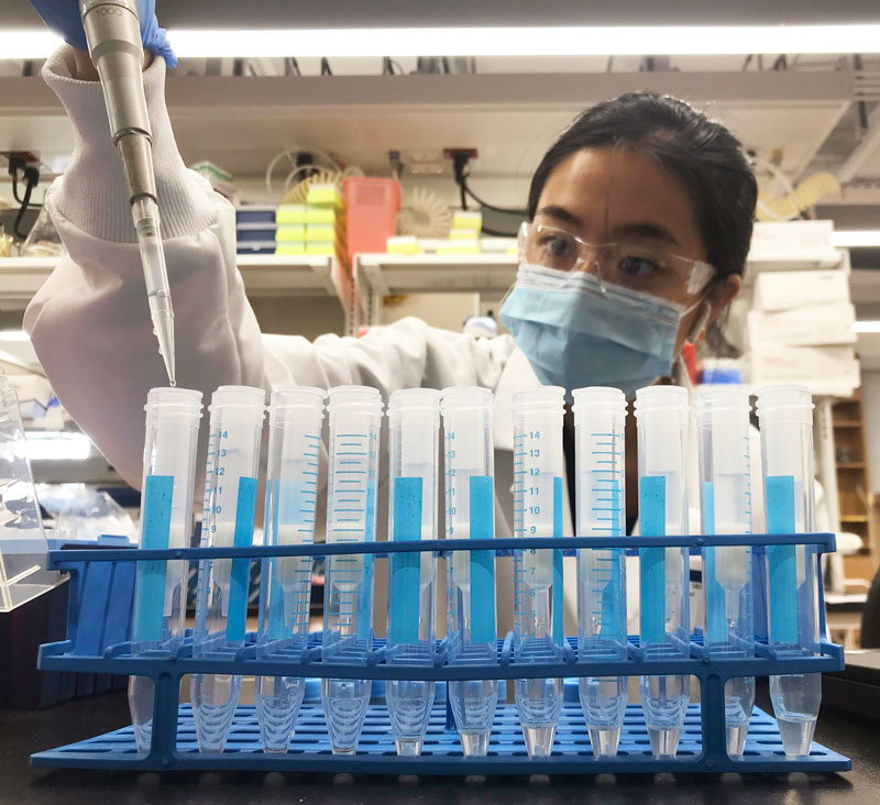
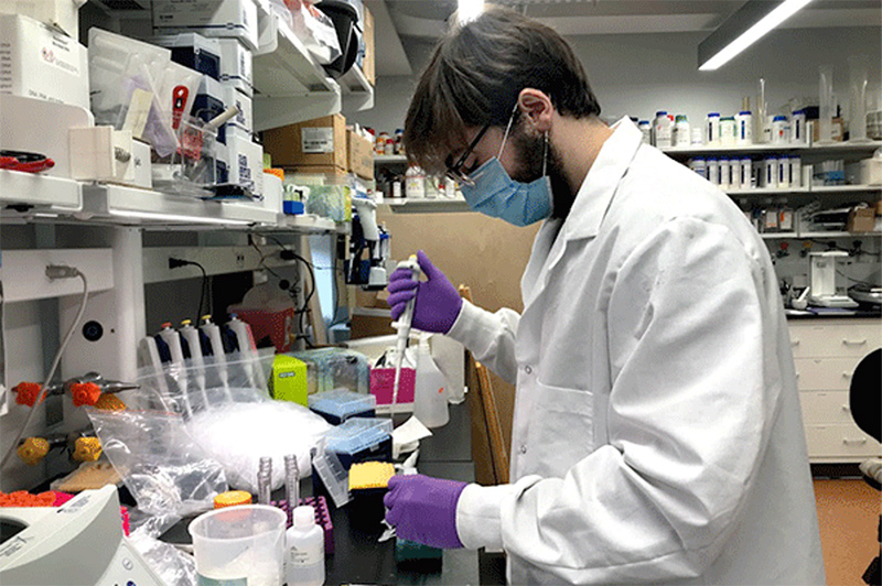
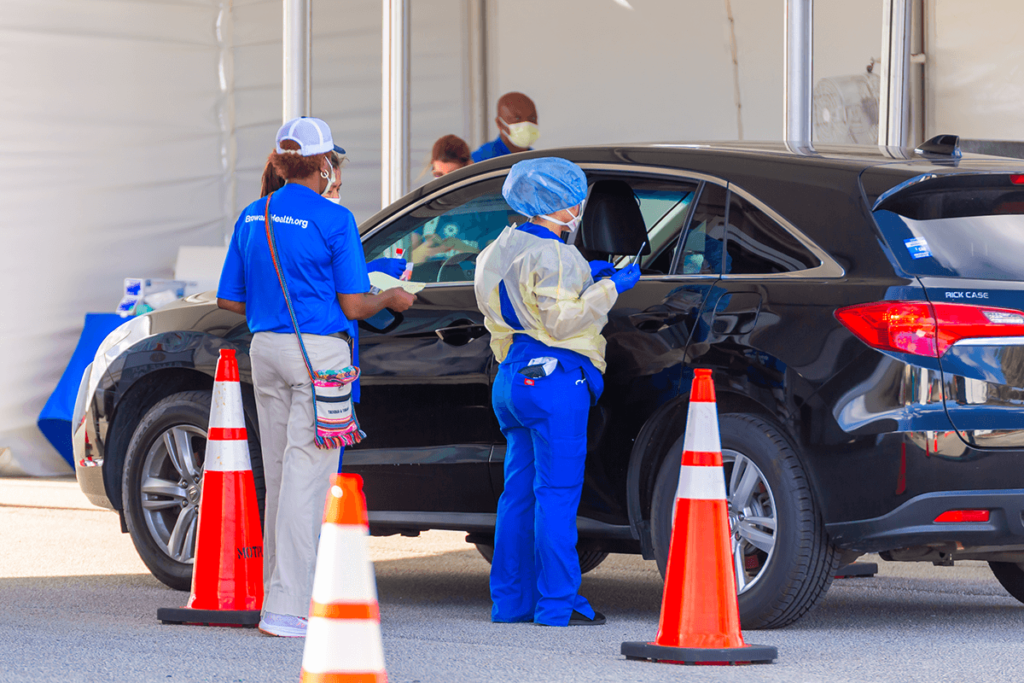
RNA Extraction for COVID-19 Testing

Once the sample arrives in the lab, the RNA needs to be purified from the sample with an extraction kit manually or with an automated liquid handler.

Shortage of RNA Extraction Kits
Just like swabs, there is a dramatic shortage of the reagents required to purify and analyze the viral samples for COVID-19 testing. Once the patient sample arrives in the lab, the RNA needs to be extracted. This process requires a series of chemical buffers that lyse, bind, and purify the viral RNA. Some labs make their own reagents to extract RNA, but most use commercial kits that take hours off the processing time.

With the increasing demand for COVID-19 testing, many labs are struggling to find reagents from their suppliers that are required to process the test. This then delays test results and increases the risk of further spread of the virus as patients may assume the delayed result means they are negative for COVID-19.

The situation has gotten so bad that some scientists have taken to twitter to request donations from other scientific labs. Since the RNA extraction kits are used in a wide range of applications, there were several labs, companies, and research institutes that were not analyzing COVID-19 that could help offset the critically low level of extraction kits.

As one of the largest manufacturers of RNA extraction kits, Zymo Research has increased their manufacturing capabilities to help support the massive need for additional reagents. They will be producing enough of their kits to enable millions of coronavirus tests each month.
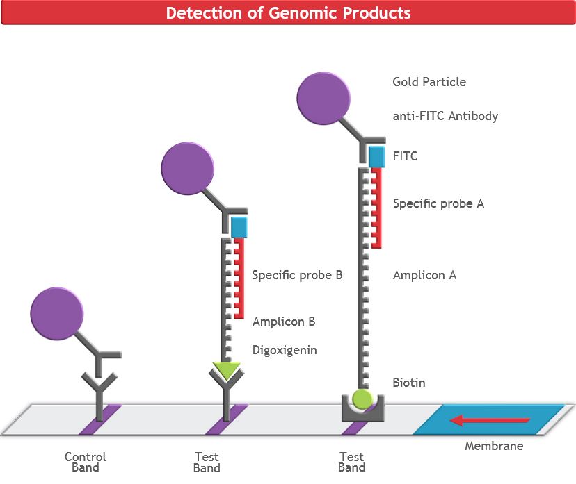
COVID-19 Detection
The purified samples contain the host RNA and any COVID-19 RNA (if present). To detect the viral RNA in the sample, a molecular biology technique called real-time reverse transcription polymerase chain reaction(rRT-PCR). This system amplifies the small amount of viral COVID-19 RNA present to thousands of copies, so that may be detected. This  makes this assay extremely sensitive.

If there is no virus present within the sample, there will be nothing for the rRT-PCR to amplify and the machine will show a negative result.  

Shortage of Reagents
Like with all other supplies in the testing workflow, the detection reagents for the rRT-PCR are also in short supply. There are shortages of the detection buffers, dyes, and enzymes will that dramatically slowdown the time to get results to patients.
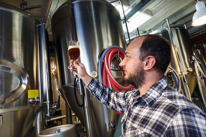
Scaling Up

Typically, when a lab begins to process a high number of samples they turn to automated solutions and move away from processing samples by hand. This is done by robotic platforms that greatly increase the throughput of a lab, allowing hundreds or even thousands of samples to be processed in a single day.

Stalled Implementation of Automation

As the testing demand for COVID-19 has grown, it has strained processing labs, and many of these laboratories are looking toward high-throughput automated workflows to keep up with the flood of patient samples that need to be processed. However, these automated workflows usually require lengthy amounts of time before a lab can fully utilize the machine because it has to be set up appropriately.

First, the machine needs to be delivered and installed. Since these are large and sensitive pieces of equipment, this process can take up to a week. Once the robot has been set up, every movement of the robot has to be coded in (or scripted) so that it can manipulate the samples appropriately and isolate the viral RNA. Just like coding an app on your phone, the scripting process is very detailed and time-consuming. Every movement of the robot has to be detailed and entered into the machine: the volume of reagent added at each step, where the reagent is dispensed, how many times the sample should be mixed, and more. Once these commands are correctly entered into the liquid handler, the script has to be tested and then optimized as needed.

From here, the script needs to be validated with test samples to ensure the device can function properly. Once it has been validated, the lab can finally begin processing patient samples on the machine.

This process is time-consuming and arduous for many labs and can take several weeks to complete from the time the device is purchased to the first patient samples being processed on it. In the COVID-19 era, these delays simply hold back labs from testing as many samples as they need to.

The best solution to avoid these delays in implementing automation is to use an automated liquid handler that comes preloaded with scripts that have already been configured and optimized for viral RNA extraction, like the DreamPrep NAP. This device delivers a load-and-go system with a user-friendly interface that dramatically decreases the amount of time needed to begin processing samples. With this device, labs can quickly and easily scale up their operations to accommodate testing for a much higher number of COVID-19 samples.


In the world of biology, this workflow is not very complicated. Theoretically, this workflow could function seamlessly with samples flowing in from patients and being processed by the lab with results delivered in 1-2 days. However, the global pandemic has placed a severe strain on the entire testing workflow. Just as with hospitals being overburdened and reaching capacity, testing labs around the world are feeling this same effect. Because of this, there is a severe delay in the implementation and expansion of critical testing services, which adds days to the processing time.

This strain on the testing system is unlike anything the field has ever seen. The industry needs rapid implementation of new solutions to combat the virus from all angles.

With scientists around the world working feverishly to expand production, implement new testing workflows, and streamline processes, the United States is slowly ramping up the amount of tests that can be administered. However, none of these tactics will be effective without the help of the public. Just as the public can flatten the curve for hospitals by staying home, that same effect applies to testing laboratories. With most of the public staying home, they are already helping to resolve the testing bottleneck by decreasing the number of samples that need to be processed. As the world comes together to fight the virus, every single person has a part to play.

See related blogs:

How To Increase Plasmid Yield

How To Increase Plasmid Yield

Sometimes, plasmid purification just doesn’t go according to plan. Whether a vector is kept at low copy number, low culture density, or culture overgrowth, this guide will help you navigate your purification woes and determine the best way to boost your plasmid yields from E. coli cultures.

Increase the Amount of Culture Processed

Sometimes the simplest way for how to increase plasmid yields is to just input more raw material. While this is simple enough in theory, there are a few considerations to observe before you start adding more culture to your plasmid preps. Most plasmid prep kits have limitations on the amount of culture they can process, and these limitations will vary based on the copy number of your plasmid as well.

The efficacy of column-based plasmid purification doesn’t just depend on how much plasmid is loaded onto the column, but also the total amount of biomass being loaded as well. If you want to scale up your plasmid purification, try using a kit designed for high inputs of culture, such the ZymoPURE II Midipreps and Maxipreps, which can process more culture.

Increasing the amount of culture processed leads to higher total yield of plasmid DNA if kit parameters are adjusted accordingly to accommodate higher biomass.

Optimize Your Bacteria

Sometimes particular E. coli strains are sub-optimal for plasmid extraction. If you are experiencing low yields for your plasmid prep, double check that the strain you’re using is best for plasmid propagation. Some strains are more optimized for protein expression than for efficient DNA replication. Others have unwanted byproducts, such as carbohydrates or endonucleases, which can co-purify with plasmids. When possible, stick to tried and tested strains like E. coli DH5α which contain mutations to lack certain endonucleases and increase plasmid stability. Thus, these modified E. coli strains are used as workhorses for molecular cloning and plasmid production.

Use Optimal Growth Conditions

Never inoculate culture straight from your bacterial stock. Always start with a single colony that is then grown as a starter culture. This ensures that your culture is derived from the same genotype and is not a mix of different colonies which may not have the same characteristics.

Also, be sure to double check your growth conditions. While many guides will recommend 12-16 hour cultures, every E. coli strain is slightly different in its growth rate and final density. Some require different temperatures, longer incubation times, faster shaking speeds, or more oxygenation.

For example, the maximum culture volume should not exceed 1/5 the total volume of the growth flask or alternatively, growth in baffled flasks can be used to increase aeration, and thus, culture growth. Also, take note of the shaking speed of the culture, with 200-250 rpm typical.

Additionally, total plasmid yield can vary depending on the type of culture broth used, typically Luria-Bertani broth (LB) or Terrific Broth (TB). Don’t be afraid to experiment with your growth conditions to see which gives you the best plasmid yield.

Purification from cultures grown in highly enriched media such as Terrific Broth (TB) yield higher amounts of plasmid DNA per ml of culture than standard Luria-Bertani Broth (LB).

Optimize Selective Pressure and Yield

There are a couple ways to ensure high yield plasmid preparations from E. coli cultures by using antibiotics for selective pressure.

The first is the antibiotic selection required for any molecular cloning experiment. It’s imperative to ensure the correct amount of antibiotic is present in your culture, otherwise the lack of selection pressure will cause your culture to start to dilute out the plasmid during cell division.

The second method uses chloramphenicol, an antibiotic that halts protein synthesis and decouples it from plasmid replication, when culturing strains containing a plasmid with a relaxed origin of replication. Chloramphenicol treatment can stop protein production but allow the E. coli  to continue to “amplify” the plasmids, resulting in increased yields during plasmid purification1.

One method for plasmid amplification uses an inhibitory amount of chloramphenicol (170 µg/ml) added to a culture, which is then incubated further until plasmid purification (typically the next day)2. A variation of this method that reports higher plasmid yield uses lower amounts of chloramphenicol (10-20 µg/ml) added to exponentially growing cells that are subsequently incubated overnight prior to plasmid purification3. Alternatively, another study demonstrated increased plasmid yield by growth in the presence of sub-inhibitory concentrations of chloramphenicol (3-5 µg/ml) from the time of culture inoculation until plasmid was harvested the next day4. Note that these treatments only work for chloramphenicol-sensitive cells and plasmids that do not encode for chloramphenicol resistance.

Bringing It Full Circle

Next time you’re stumped about your plasmid purification, apply these tips on how to increase your plasmid yields and optimize your experiment. Whether it be increasing the volume, changing your growth conditions, adding more selective pressure, or using the best isolation technologies, there is always optimization to be done to increase your plasmid yields!

Learn how to collect high purity plasmid DNA:

References:

1. Ausubel, FM, et al. Current Protocols in Molecular Biology, (2003)
2. Sambrook, J, Fritsch EF, Maniatis T, Molecular Cloning: a laboratory manual, 2nd edition. Cold Springs Harbour Laboratory Press, Cold Springs Harbour, New York, 1989
3. Frenkel L, Bremer H, Increased Amplification of Plasmids pBR322 and PBR327 by Low Concentrations of Chloramphenicol, DNA 5, num 6, 539-544 (1986)
4. Begbie S, et al., The Effects of Sub-Inhibitory Levels of Chloramphenicol on pBR322 Plasmid Copy Number in Escherichia coli DH5a Cells, Journal of Experimental Microbiology and Immunology 7, 82-88 (2005)
5. https://www.zymoresearch.com/


What Are Transformation, Transfection & Transduction?

What Are Transformation, Transfection & Transduction?

One of the pillars of modern day molecular biology uses techniques to manipulate DNA sequences (such as plasmids, knockout gene constructs, etc.) and introduce them into a host cell to test their effects. However, getting the DNA into cells can take different routes. Those unfamiliar with the field may be wondering “what is plasmid transduction?” Or have heard the terms transformation, transfection, and transduction, but are uncertain as to the differences and similarities between these techniques. Although these terms have some overlap, and so their usage is often confusing or incorrect.

What Is Plasmid Transformation?

Transformation is, simply put, the process of altering a cell’s genetic code through the uptake of foreign DNA from the environment. Plasmid transformation is used to describe the (non-viral) horizontal gene transfer of plasmids between bacteria. While transformation likely happens in the natural world, scientists have harnessed this process to their own ends, enabling replication of lab-manipulated plasmids and expression of desired recombinant DNA sequences.

The process is relatively simple; scientists make the membranes of bacterial cells permeable to DNA either through chemical means or via electrical stimulation. These cells, now termed ‘competent cells,’ will readily uptake plasmid DNA from their surroundings. Once the DNA molecule of interest is introduced to these competent cells, the bacteria have now been plasmid transformed. The transformed cells then can be selected from the untransformed cells by inclusion of an antibiotic to kill off the untransformed cells. Typically, this occurs as the plasmid will express an antibiotic resistance gene to protect the transformed cells and ensure maintenance of the plasmid over time and cell divisions. In the process, many replicons of the plasmid will be created and passed to daughter cells.

What Is Plasmid Transfection?

Transfection is a type of plasmid transformation, typically that of animal cells, instead of bacteria. This process is a bit more complicated than your run-of-the-mill transformation, as many lab-cultured eukaryotic cells do not natively uptake and replicate foreign DNA. Still, scientists have discovered many ways in which plasmids and other foreign DNA can be introduced to cells.

Much like methods for bacteria, there are both chemical and physical methods of transfection produce transient holes in the cell membrane and get uptake of foreign DNA. These methods work similarly to the those outlined for bacterial transformation, as they all are designed to make the cell membrane more permeable. The method by which they do so is different from bacteria, though, instead using cationic lipids, micelles, lasers, or even particle guns. These methods have their pros and cons, but ultimately will depend on the resources available and the preference of the researcher.

What Is Transduction?

The final prominent method, transduction, is unique from the other two methods. Transduction is the process of using a virus to mediate the delivery of DNA fragments or plasmids into a cell, either prokaryotic or eukaryotic. This technique harnesses the natural function of viruses to inject DNA into the infected host, but with a twist. Scientists can modify the viral nucleic acids to contain specific DNA sequences of interest. There are many different types of viruses that can be manipulated to introduce recombinant nucleic acids into host cells. For example, bacteriophage introduce DNA into bacteria, and lentiviruses or adenoviruses into human cells. Using these modified viruses, researchers incorporate foreign DNA into the host genome (such as using lentiviruses or bacteriophage) or transiently express desired recombinant nucleic acids (such as using adenoviruses).

In order to perform a transduction, you need a cell-line of interest and a virus that infects that cell line. This method can be more difficult than the other methods discussed here, since the virus must be grown and maintained in culture, sometimes needs to be modified to be non-infectious to humans, and the DNA of interest must be packaged into the viral particle before infection of new host cells can occur. Despite the challenges to overcome, viral transduction is an excellent way to perform stable, long term transformations and transfections in the lab environment.

Learn how to collect high purity plasmid DNA: