Home >Protein Sample Preparation and Cell Fractionation >Minute™ Mitochondria Isolation Kit for Mammalian Cells and Tissues (50 Preps)
Minute™ Mitochondria Isolation Kit for Mammalian Cells and Tissues (50 Preps)
Manual & Protocol (PDF) | Material Safety Data Sheets (MSDS)(PDF)
View All Related Products
Description
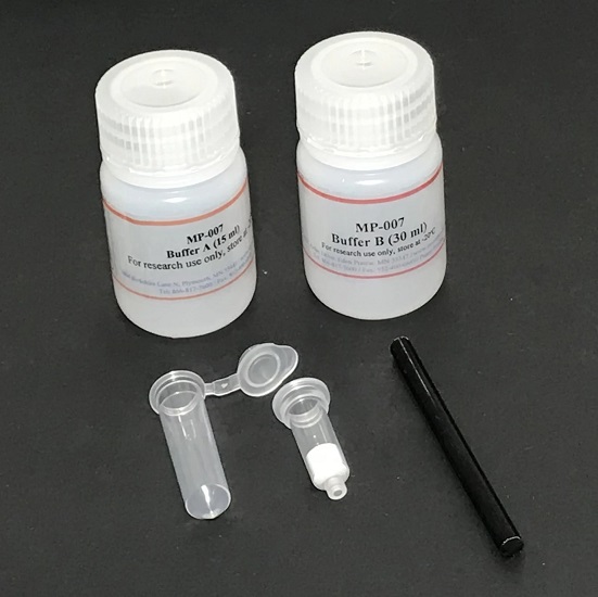
Minute™ Mitochondria Isolation Kit for Mammalian Cells and Tissues is composed of optimized buffers and protein extraction filter cartridges with 2.0 ml collection tubes. The kit is designed to rapidly isolate native mitochondria proteins from cultured mammalian cells or tissues. Due to the use of protein extraction filter cartridges, the protein isolation procedure is simple and provides a high yield. The procedure can be completed in less than 30 minutes. Unlike many commercial mitochondria preparation kits, this kit offers a wide range of starting cells and isolated mitochondria. The buffers are detergent and EDTA free. A Dounce homogenizer or a tissue blender is not needed.
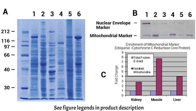
Figure 1. Enrichment of Mitochondrial Marker (Ubiquinol-Cytochrome C Reductase Core Protein) with Minute™ Mitochondria Isolation Kit.
A. SDS-PAGE profiles of total protein extract vs. isolated mitochondrial proteins. Lanes 1, 3, 5, total proteins from isolated mouse kidney cells, mouse skeletal muscle and liver tissues, respectively. Lanes 2, 4, 6, isolated mitochondrial proteins from mouse kidney cells, mouse skeletal muscle and liver tissues, respectively.
B. Western blotting. Proteins in A were transferred to nitrocellulose membranes and probed with anti-ubiquinol-cytochrome C reductase core protein (ab96333, abcam, Cambridge, MA) and anti-lamin B1 (ab16048, a nuclear envelope marker protein. abcam, Cambridge, MA).
C. Densitometry measurement of mitochondrial marker signals.
Kit Includes
| Items | Quantity |
| Buffer A | 20 ml |
| Buffer B | 8 ml |
| Protein Extraction Filter Cartridges | 50 Units |
| Collection Tubes with caps | 50 Units |
| Plastic Rods | 2 Units |
References
1. Gou, R. et al. (2014). Resveratrol suppresses oxidized low-density lipoprotein-induced macrophage apoptosis through inhibition of intracellular reactive oxygen species generation, lox-1 and the p38 MAPK pathway. Cellular Physiology and Biochemistry. 34:603-616.
2. Hu, H., Zhu, W., Qin, J., Chen, M., Gong, L., Li, L., ... & Ye, D. (2016). Acetylation of PGK1 Promotes Liver Cancer Cell Proliferation and Tumorigenesis. Hepatology.
3. Gao, G., Wang, Z., Lu, L., Duan, C., Wang, X., & Yang, H. (2017). Morphological analysis of mitochondria for evaluating the toxicity of α-synuclein in transgenic mice and isolated preparations by atomic force microscopy. Biomedicine & Pharmacotherapy.
4. Zhang, L., Liu, J., Zhou, F., Wang, W., & Chen, N. (2018). PGC-1α ameliorates kidney fibrosis in mice with diabetic kidney disease through an antioxidative mechanism. Molecular medicine reports.
5. Wu, Y., Gao, W. N., Xue, Y. N., Zhang, L. C., Zhang, J. J., Lu, S. Y., ... & Sun, L. K. (2018). SIRT3 aggravates metformin-induced energy stress and apoptosis in ovarian cancer cells. Experimental Cell Research.
6. Shen, L., Sun, B., Sheng, J., Yu, S., Li, Y., Xu, H., ... & Sun, L. (2018). PGC1α promotes cisplatin resistance in human ovarian carcinoma cells through upregulation of mitochondrial biogenesis. International Journal of Oncology.
7.Han, W., Cao, F., Gao, X. J., Wang, H. B., Chen, F., Cai, S. J., ... & Ding, H. Z. ZIC1 acts a tumor suppressor in breast cancer by targeting survivin. International Journal of Oncology.
8. Ouyang, J., Zeng, Z., Fang, H., Li, F., Zhang, X., & Tan, W. (2019). SIRT3 inactivation promotes acute kidney injury through elevated acetylation of SOD2 and p53. Journal of Surgical Research, 233, 221-230.
9. Zhou, K., Yao, Y. L., He, Z. C., Chen, C., Zhang, X. N., Yang, K. D., ... & Niu, Q. (2018). VDAC2 interacts with PFKP to regulate glucose metabolism and phenotypic reprogramming of glioma stem cells. Cell Death & Disease, 9(10), 988.
10. Xue, Y. N., Yu, B. B., Li, J. L., Guo, R., Zhang, L. C., Sun, L. K., ... & Li, Y. (2018). Zinc and p53 Disrupt Mitochondrial Binding of HK2 by Phosphorylating VDAC1. Experimental Cell Research.
11. Chen, X. Y., Ren, H. H., Wang, D., Chen, Y., Qu, C. J., Pan, Z. H., ... & Li, D. F. (2019). Isoliquiritigenin Induces Mitochondrial Dysfunction and Apoptosis by Inhibiting mitoNEET in a Reactive Oxygen Species-Dependent Manner in A375 Human Melanoma Cells. Oxidative Medicine and Cellular Longevity, 2019.
12. Ma, J., Chen, L., He, X. X., Wang, Y. J., Yu, H. L., He, Z. X., ... & Zhu, X. J. (2019). Functional prediction and characterization of Dip2 gene in mice. Cell biology international.
13. Cai, M., He, P., & Fang, D. L. Hypoxia‑induced mitochondrial translocation of DNM1L increases mitochondrial fission and triggers mPTP opening in HCC cells via activation of HK2. Oncology Reports.
14. Guo, Z., Song, T., Xue, Z., Liu, P., Zhang, M., Zhang, X., & Zhang, Z. (2019). Using CETSA assay and a mathematical model to reveal dual Bcl-2/Mcl-1 inhibition and on-target mechanism for ABT-199 and S1. European Journal of Pharmaceutical Sciences, 105105.
15.Yue, J., Shen, Y., Liang, L., Cong, L., Guan, X., Li, Z., ... & Xu, W. In situ and ex situ surface‐enhanced Raman spectroscopy (SERS) analysis of cell mitochondria. Journal of Raman Spectroscopy.

Post Reviews
 We're here to help
Get expert recommendations for common problems or connect directly with an on staff expert for technical assistance related to applications, equipment and general product use.
Contact Tech Support
 High Quality Guaranteed Product
Our products such as Elisa, Antibodies, Proteins, Peptides and sequencing kits are covered by Biolinkk quality warranty and will work as described in datasheet, a free replacement or money back is guaranteed if does not perform according to datasheet.
Learn More