
Antibody Testing Data:
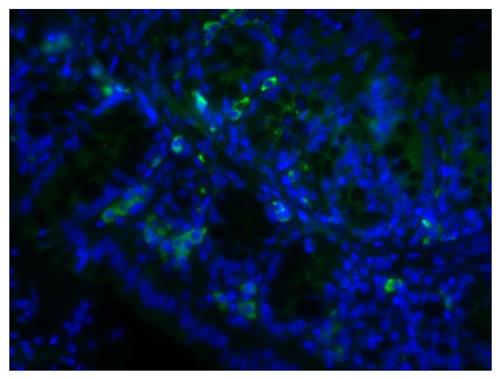
Paraffin embedded mouse small intestine tissue section was blocked with Normal Rabbit Serum (SB Cat. No. 0040-01) and stained with Goat Anti-Mouse IgA-UNLB (SB Cat. No. 1040-01) followed by Rabbit Anti-Goat IgG(H+L), Human SP ads-BIOT (SB Cat. No. 6164-08), Streptavidin-FITC (SB Cat. No. 7100-02), DAPI, and mounted with Fluoromount-G® Anti-Fade (SB Cat. No. 0100-35).
Product Details
Product Specifications |
|
|---|---|
|
Name: |
Rabbit Anti-Goat IgG(H+L), Human SP ads-BIOT |
| Clone: |
Polyclonal |
|
Isotype: |
Rabbit IgG (Isotype Control: Rabbit IgG-BIOT) |
|
Specificity: |
Reacts with the heavy and light chains of goat IgG |
| Format/Conjugate: |
BIOT (Biotin) |
| Buffer Formulation: |
Phosphate buffered saline containing 0.1% sodium azide |
|
Concentration: |
0.5 mg/mL |
|
Volume: |
2.0 mL |
|
Storage & Handling: |
2-8°C blue Please refer to product specific SDS. |
| RRID: |
AB_2796239 |
|
Recommended Dilutions: |
Please refer to product specific Technical Guide. |
|
Applications: |
Quality tested applications for relevant formats include - |
|
More information: |
Source: Pooled antisera from rabbits hyperimmunized with goat IgG |
*For Research Use Only. Not for use in diagnostic procedures. Not for resale without express authorization.
Documents
Technical Guide
 Technical Guide: Rabbit Anti-Goat IgG(H+L), Human SP ads-BIOT
Safety Data Sheets (SDS)
SDS: Rabbit Anti-Goat IgG(H+L), Human SP ads-BIOT References
1. Boxx GM, Kozel TR, Nishiya CT, Zhang MX. Influence of mannan and glucan on complement activation and C3 binding by Candida albicans. Infect Immun. 2010;78:1250-9. (ELISA)
2. Fu S, Gong F, Xie C, Zhu W, Wang W, Shen H, et al. S100A12 on circulating endothelial cells surface in children with Kawasaki disease. Pediatr Res. 2010;68:165-8. (FC)
3. Rezaee F, Rellick SL, Piedimonte G, Akers SM, O'Leary HA, Martin K, et al. Neurotrophins regulate bone marrow stromal cell IL-6 expression through the MAPK pathway. PLoS One. 2010;5(3):e9690. (FC)
4. Sekiya I, Ojima M, Suzuki S, Yamaga M, Horie M, Koga H, et al. Human mesenchymal stem cells in synovial fluid increase in the knee with degenerated cartilage and osteoarthritis. J Orthop Res. 2012;30:943-9. (FC)
5. Gong F, Zhang Y, Xie C, Zhu W, Wang W, Fu S, et al. Expression of receptor for advanced glycation end products (RAGE) on the surface of circulating endothelial cells is upregulated in Kawasaki disease. Pediatr Res. 2012;71:720-4. (FC)
6. Hegde M, Corder A, Chow KK, Mukherjee M, Ashoori A, Kew Y, et al. Combinational targeting offsets antigen escape and enhances effector functions of adoptively transferred T cells in glioblastoma. Mol Ther. 2013;21:2087-101. (FC)
7. Kreft KL, Verbraak E, Wierenga-Wolf AF, van Meurs M, Oostra BA, Laman JD, et al. The IL-7Rα pathway is quantitatively and functionally altered in CD8 T cells in multiple sclerosis. J Immunol. 2012;188:1874-83. (IHC-FS)
8. Crijns AP, de Graeff P, Geerts D, ten Hoor KA, Hollema H, van der Sluis T, et al. MEIS and PBX homeobox proteins in ovarian cancer. Eur J Cancer. 2007;43:2495-505. (IHC-PS)
9. Ando K, Brion J, Stygelbout V, Suain V, Authelet M, Dedecker R, et al. Clathrin adaptor CALM/PICALM is associated with neurofibrillary tangles and is cleaved in Alzheimer's brains. Acta Neuropathol. 2013;125:861-78. (IHC-PS)
10. Ahmed L, Nalwoga H, Arnes JB, Wabinga H, Micklem DR, Akslen LA. Increased tumor cell expression of Axl is a marker of aggressive features in breast cancer among African women. APMIS. 2015 May 25. doi: 10.1111/apm.12403. [Epub ahead of print]. (IHC-PS)
11. Hosper NA, van den Berg PP, de Rond S, Popa ER, Wilmer MJ, Masereeuw R, et al. Epithelial-to-mesenchymal transition in fibrosis: collagen type I expression is highly upregulated after EMT, but does not contribute to collagen deposition. Exp Cell Res. 2013;319:3000-9. (ICC)
12. Rayes J, Roumenina LT, Dimitrov JD, Repessé Y, Ing M, Christophe O, et al. The interaction between factor H and VWF increases factor H cofactor activity and regulates VWF prothrombotic status. Blood. 2014;123:121-5. (WB)
13. Wu X, Sagave J, Rutkovskiy A, Haugen F, Baysa A, Nygård S, et al. Expression of bone morphogenetic protein 4 and its receptors in the remodeling heart. Life Sci. 2014;97:145-54. (WB)
14. Matsebatlela TM, Anderson AL, Gallicchio VS, Elford H, Rice CD. 3,4-Dihydroxy-benzohydroxamic acid (Didox) suppresses pro-inflammatory profiles and oxidative stress in TLR4-activated RAW264.7 murine macrophages. Chem Biol Interact. 2015;233:95-105. (WB)
 We're here to help
Get expert recommendations for common problems or connect directly with an on staff expert for technical assistance related to applications, equipment and general product use.
Contact Tech Support
 High Quality Guaranteed Product
Our products such as Elisa, Antibodies, Proteins, Peptides are covered by Biolinkk quality warranty and will work as described in datasheet, a free replacement or money back is guaranteed if does not perform according to datasheet.
Learn More