Minute™ High-Efficiency Exosome Precipitation Reagent

Home / Articles posted byadmin

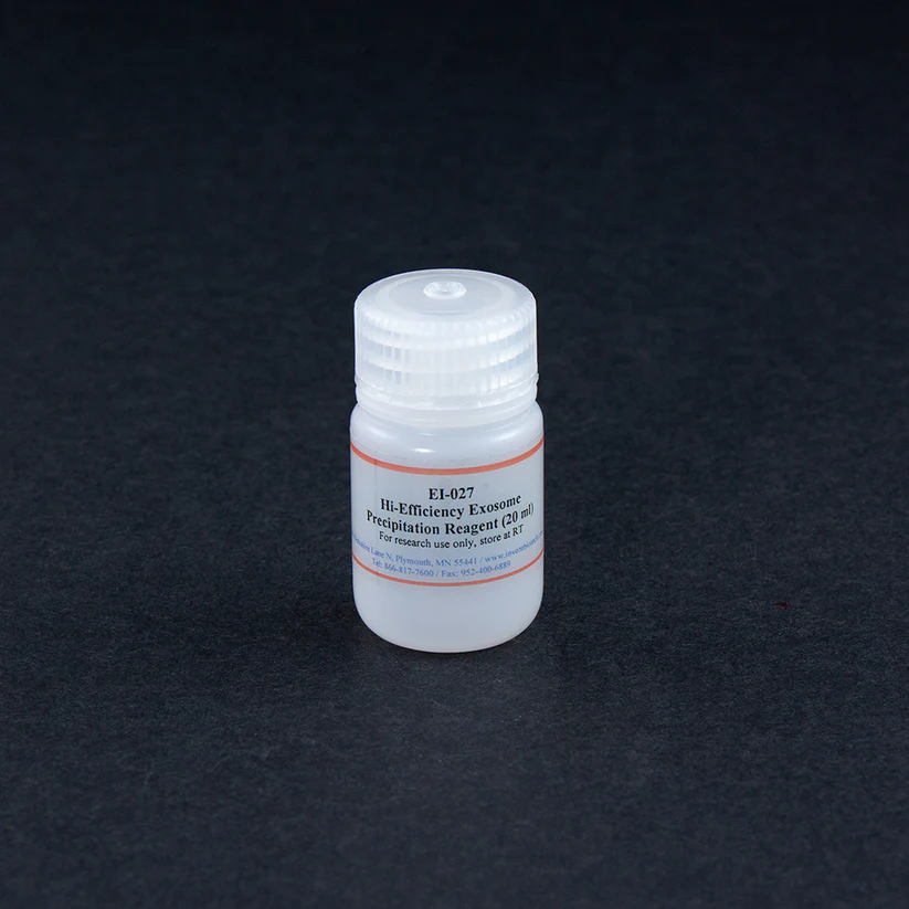
Minute™ High-Efficiency Exosome Precipitation Reagent (20 ml)

Cat# : EI-027

Description

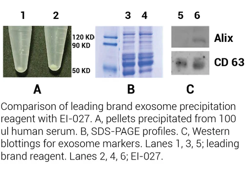
Exosomes are vesicles secreted by cells. They are present in a variety of body fluids such as serum, ascites, spinal cord fluid, urine, and saliva. Cultured cells also secrete significant number of exosomes. The size distribution of exosome is ranging from 30-120 nm. The biological function of exosomes is believed to serve as intercellular messengers. Filtration and ultracentrifugation are classical ways of isolating and enriching exosomes. This is a tedious process and requires special equipment. Another common way for enrichment of exosome is through precipitation. Currently major commercial kits are PEG-based. EI-027 is designed to precipitate total exosomes from biofluids using a high efficacy, non-PEG based reagent for exosome precipitation. This kit is suitable for routine biofluid samples using the same reagent and similar protocol.

Kit includes:

Item

Quantity

Exosome Precipitation Reagent

20 ml

References (11)
    1. Bai, H., Pan, Y., Qi, L., Liu, L., Zhao, X., Dong, H., … & Wang, X. (2018). Development a hydrazide-functionalized thermosensitive polymer based homogeneous system for highly efficient N-glycoprotein/glycopeptide enrichment from human plasma exosome. Talanta.
    2. Jiao, F., Xia, C., Zhao, Y., Ying, W., Xie, Y., Guan, X., … & Qian, X. (2018). A novel strategy for facile serum exosome isolation based on specific interactions between phospholipid bilayers and TiO2. Chemical Science.
    3. Tan, Y., Nie, W., Chen, C., He, X., Xu, Y., Ma, X., … & Wang, W. (2019). Mesenchymal stem cells alleviate hypoxia-induced oxidative stress and enhance the pro-survival pathways in porcine islets. Experimental Biology and Medicine, 1535370219844472.
    4. Cheng, P., Feng, F., Yang, H., Jin, S., Lai, C., Wang, Y., & Bi, J. (2020). Detection and significance of exosomal mRNA expression profiles in the cerebrospinal fluid of patients with meningeal carcinomatosis. Journal of Molecular Neuroscience, 1-14.
    5. Lu, T., Zhang, Z., Zhang, J., Pan, X., Zhu, X., Wang, X., … & Yan, M. (2022). CD73 in small extracellular vesicles derived from HNSCC defines tumour‐associated immunosuppression mediated by macrophages in the microenvironment. Journal of Extracellular Vesicles11(5), e12218.
    6. Theel, E. K., & Schwaminger, S. P. (2022). Microfluidic Approaches for Affinity-Based Exosome Separation. International Journal of Molecular Sciences23(16), 9004.
    7. Omrani, M., Beyrampour-Basmenj, H., Jahanban-Esfahlan, R., Talebi, M., Raeisi, M., Serej, Z. A., … & Ebrahimi-Kalan, A. (2023). Global trend in exosome isolation and application: an update concept in management of diseases. Molecular and Cellular Biochemistry, 1-13.
    8. Omrani, M., Beyrampour-Basmenj, H., Jahanban-Esfahlan, R., Talebi, M., Raeisi, M., Serej, Z. A., … & Ebrahimi-Kalan, A. (2023). Global trend in exosome isolation and application: an update concept in management of diseases. Molecular and Cellular Biochemistry, 1-13.
    9. Cheng, C., Wang, P., Yang, Y., Du, X., Xia, H., Liu, J., … & Liu, Q. (2023). Smoking‐Induced M2‐TAMs, via circEML4 in EVs, Promote the Progression of NSCLC through ALKBH5‐Regulated m6A Modification of SOCS2 in NSCLC Cells. Advanced Science, 2300953.
    10. Chen, G., Pu, G., Wang, L., Li, Y., Liu, T., Li, H., … & Luo, X. (2023). Cysticercus pisiformis-derived novel-miR1 targets TLR2 to inhibit the immune response in rabbits. Frontiers in Immunology14.

    11. Cheng, C., Wang, P., Yang, Y., Du, X., Xia, H., Liu, J., … & Liu, Q. (2023). Smoking‐Induced M2‐TAMs, via circEML4 in EVs, Promote the Progression of NSCLC through ALKBH5‐Regulated m6A Modification of SOCS2 in NSCLC Cells. Advanced Science, 2300953.

Minute™ Nuclear Envelope Protein Extraction Ki

Home / Articles posted byadmin

Minute™ Nuclear Envelope Protein Extraction Kit (50 Preps)

Cat# : NE-013

Description

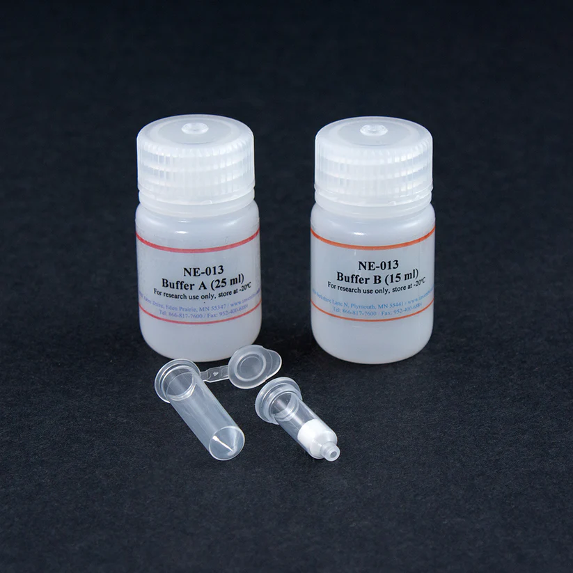
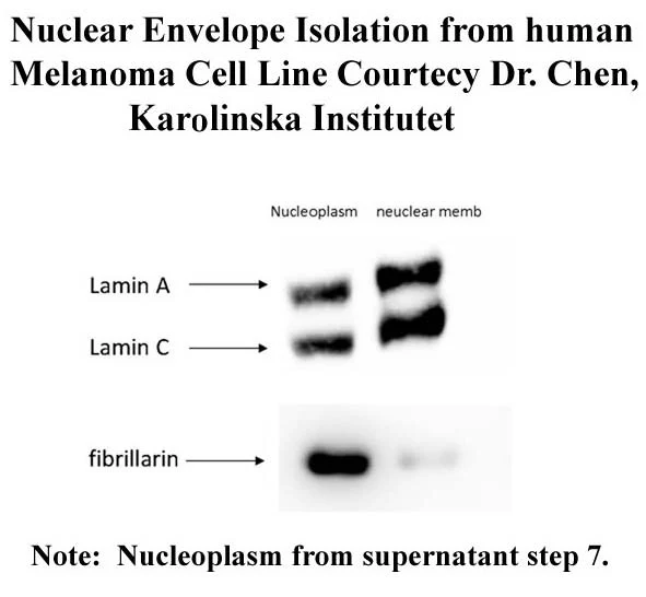
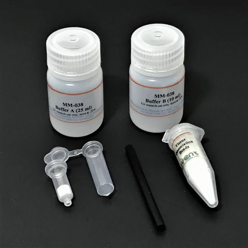
The nuclear envelope is a very complex membrane-protein system that is notoriously difficult to isolate and purify because of its connection to nuclear and cytoplasmic components. Traditional methods of nuclear envelope isolation and purification require a large amount of starting material and a long, tedious procedure. Minute™ Nuclear Envelope Protein Extraction Kit is the first commercial kit designed to rapidly isolate nuclear envelope and its associated proteins in native form without using density-gradient and ultracentrifugation. Due to the use of protein extraction filter cartridges, the nuclear envelope protein isolation is simple and user-friendly. The nuclear envelope proteins are significantly enriched in the final prep. Unlike traditional methods that require large amounts of starting cells/tissues, this kit starts with only 10-20 million cells, and the buffers are detergent and EDTA free. The procedure can be completed in less than 45 minutes with a final yield of 10-50 µg protein/sample.

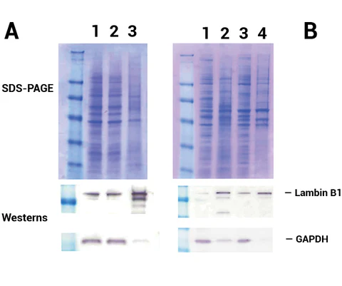
A. SDS-PAGE profiles and Western blottings of isolated mouse spleen cells. Lanes 1 and 2, total cell lysate; Lane 3, nuclear envelope.

B. SDS-PAGE profiles and Western blottings of Rate (REL-6TN) and human (MCF-7) cell lines. Lane 1, REL-6TN total cell lysate; Lane 2, REL-6TN nuclear envelope. Lane 3, MCF-7 total cell lysate; Lane 4, MCF-7 nuclear envelope. Nuclear envelope marker Lamin B1 (ab 16048, ABcam, Cambridge MA) and GAPDH (G9545, Sigma) were visualized by a color metric substrate (Bio-Rad).

Kit includes:

Items

Quantity

Buffer A

25 ml

Buffer B

15 ml

Filter Cartridges

50 units

Collection Tubes with Caps

50 units

References (7)
  1. Frias A. et al. (2016). A Switch in Akt Isoforms Is Required for Notch-Induced Snail1 Expression and Protection from Cell Death. Mol Cell Biol. 2016 Mar 15; 36(6): 913–922. Prepublished online 2015 Dec 28. Published online 2016 Mar 1. doi: 10.1128/MCB.00842-15.
  2. Selby, T. L., Biel, N., Varn, M., Patel, S., Patel, A., Hilding, L., … & Lowrey, A. J. (2019). The Epstein-Barr Virus Oncoprotein, LMP1, Regulates the Function of SENP2, a SUMO-protease. Scientific Reports9(1), 9523.
  3. Shao, Y., Li, L., Liu, L., Yang, Y., Huang, J., Ji, D., … & Sun, B. (2022). CD44/ERM/F‐actin complex mediates targeted nuclear degranulation and excessive neutrophil extracellular trap formation during sepsis. Journal of Cellular and Molecular Medicine.
  4.  Zervopoulos, S. D., Boukouris, A. E., Saleme, B., Haromy, A., Tejay, S., Sutendra, G., & Michelakis, E. D. (2022). MFN2-driven mitochondria-to-nucleus tethering allows a non-canonical nuclear entry pathway of the mitochondrial pyruvate dehydrogenase complex. Molecular Cell82(5), 1066-1077.

  5. Kovacs, M. T., Vallette, M., Wiertsema, P., Dingli, F., Loew, D., de Freitas Nader, G. P., … & Ceccaldi, R. (2023). DNA damage induces nuclear envelope rupture through ATR-mediated phosphorylation of lamin A/C. Molecular Cell.
  6. Zhou, X. Y., Lin, B., Chen, W., Cao, R. Q., Guo, Y., Said, A., … & Zhu, Y. M. (2023). The brain protection of MLKL inhibitor necrosulfonamide against focal ischemia/reperfusion injury associating with blocking the nucleus and nuclear envelope translocation of MLKL and RIP3K. Frontiers in Pharmacology14.
  7. Taga, K., & Takeuchi, H. (2024). Novel role of host protein SLC25A42 in the HIV‐1 reactivation of latent HIV‐1 provirus. Microbiology and Immunology.

Minute™ Mitochondria Isolation Kit for Mammalian Cells and Tissues

Home / Articles posted byadmin

Minute™ Mitochondria Isolation Kit for Mammalian Cells and Tissues (50 Preps)

Cat# : MP-007

Description

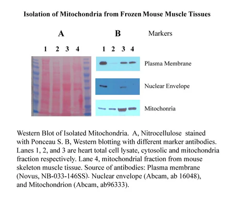
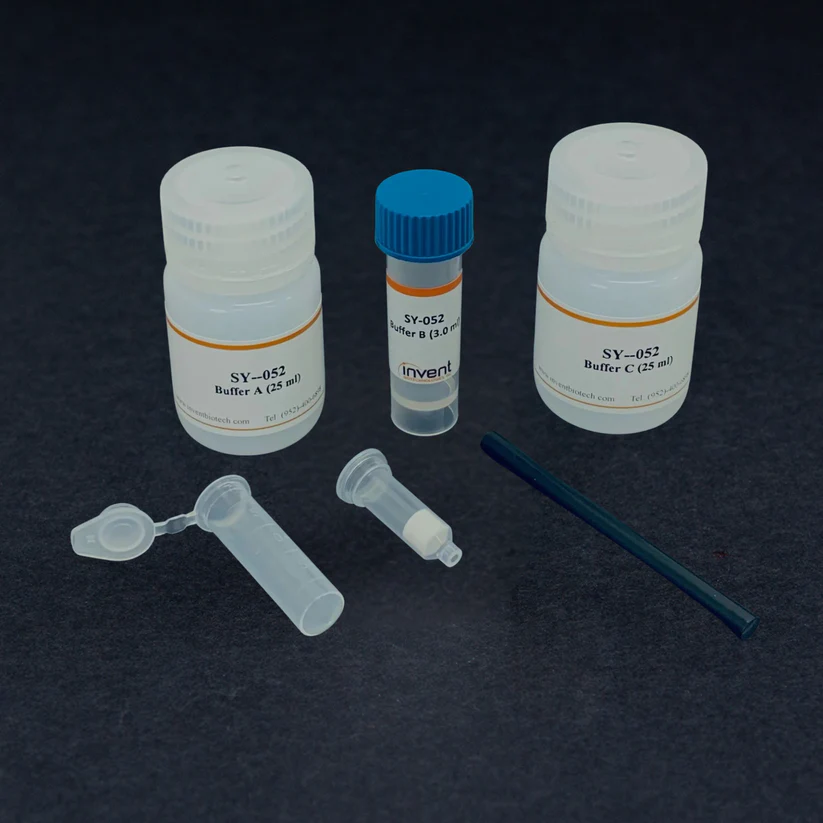
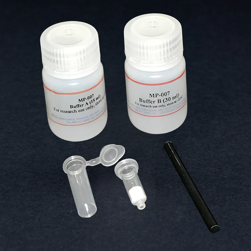
Minute™ Mitochondria Isolation Kit for Mammalian Cells and Tissues comprises optimized buffers and protein extraction filter cartridges with 2.0 ml collection tubes. The kit is designed to rapidly isolate native mitochondria proteins from cultured mammalian cells or tissues. Due to protein extraction filter cartridges, the protein isolation procedure is simple and provides a high yield. The process can be completed in less than 30 minutes. Unlike many commercial mitochondria preparation kits, this kit offers a wide range of starting cells and isolated mitochondria. The buffers are detergent and EDTA-free. A Dounce homogenizer or a tissue blender is not needed.

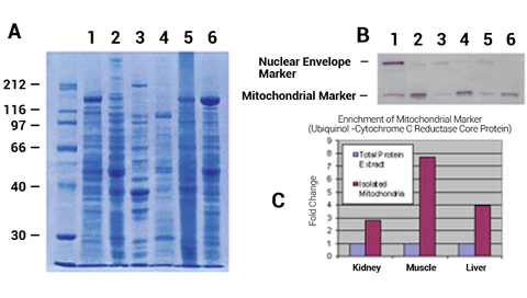
Figure 1. Enrichment of Mitochondrial Marker (Ubiquinol-Cytochrome C Reductase Core Protein) with Minute™ Mitochondria Isolation Kit. 

 A. SDS-PAGE profiles of total protein extract vs. isolated mitochondrial proteins. Lanes 1, 3, 5, total proteins from isolated mouse kidney cells, mouse skeletal muscle and liver tissues, respectively. Lanes 2, 4, 6, isolated mitochondrial proteins from mouse kidney cells, mouse skeletal muscle and liver tissues, respectively.

 B. Western blotting. Proteins in A were transferred to nitrocellulose membranes and probed with anti-ubiquinol-cytochrome C reductase core protein (ab96333, abcam, Cambridge, MA) and anti-lamin B1 (ab16048, a nuclear envelope marker protein. abcam, Cambridge, MA). 

 C. Densitometry measurement of mitochondrial marker signals.

Kit includes:

Items

Quantity

Buffer A

15 ml

Buffer B

30 ml

Protein Extraction Filter Cartridges

50 units

Collection Tubes with Caps

50 units

Plastic Rods

2 units


Minute™ Plant Cytosolic and Nuclear Protein Isolation Kit

Home / Articles posted byadmin

Minute™ Plant Cytosolic and Nuclear Protein Isolation Kit (Need 150 mg Raw Material, 20 preps)

Cat# : PF-045

Description

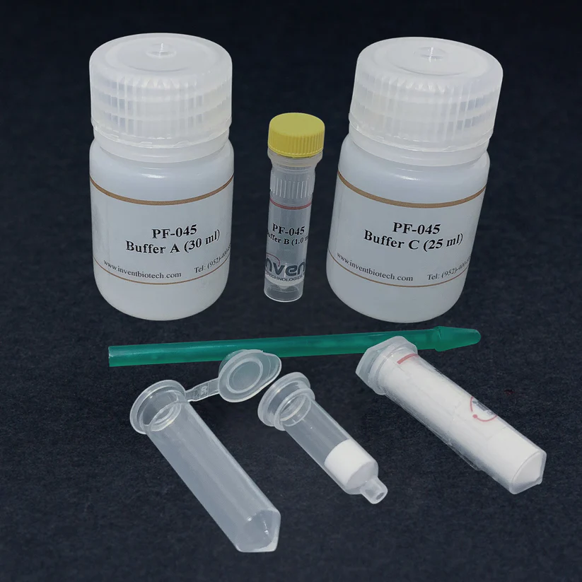
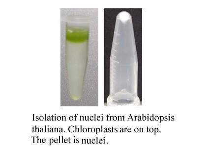
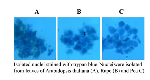
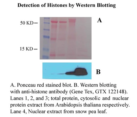
The kit is designed for rapid fractionation of fresh/frozen soft plant tissues, especially leaves. Unlike other commercial kit and Lab protocols, of which large amount of starting material (a few grams) and lengthy processing time are required, this kit only uses 100-200 mg starting material.  By using a specialized filter cartridge coupled with our proprietary buffer system, four subcellular fractions (cytosol, nuclei, chloroplasts and organelles) can be obtained in about 1 hour. The subcellular fractions can be used in numerous applications that include but not limited to protein trafficking, nuclear protein extraction and nucleic acid purification/sequencing. The highly enriched nuclei can be used for proteomics, flow cytometry analysis, DNA sequencing, and protein-protein/protein-DNA interaction studies. The subcellular fractionation protocol includes grinding tissue to release plant cells followed by disruption of the cell membrane by rapidly passing through the filter. Different subcellular fraction is obtained by differential centrifugation using only a table top centrifuge. This kit has been tested for use with many species such as Arabidopsis, Spinach, Peas, and Rape.

Kit includes:

Items

Quantity

Buffer A

30 ml

Buffer B

1 ml

Buffer C

25 ml

Filter Cartridges with Collection Tubes

20 units

Pestle for 1.5 ml Eppendorf tubes

2 units

Protein Extraction Power

2 gram


References (2)

1. Son, S., Moon, S. J., Kim, H., Lee, K. S., & Park, S. R. (2021). Identification of a novel NPR1 homolog gene, OsNH5N16, which contributes to broad-spectrum resistance in rice. Biochemical and Biophysical Research Communications549, 200-206.

2. Im, J. H., Son, S., Ko, J. H., Kim, K. H., An, C. S., & Han, K. H. (2021). Nuclear Translocation of Soybean MPK6, GmMPK6, Is Mediated by Hydrogen Peroxide in Salt Stress. Plants10(12), 2611.

Minute™ Total Lipid Raft Isolation Kit for Mammalian Cells/Tissues

Home / Articles posted byadmin

Minute™ Total Lipid Raft Isolation Kit for Mammalian Cells/Tissues (20 preps)

Cat# : LR-039

Description

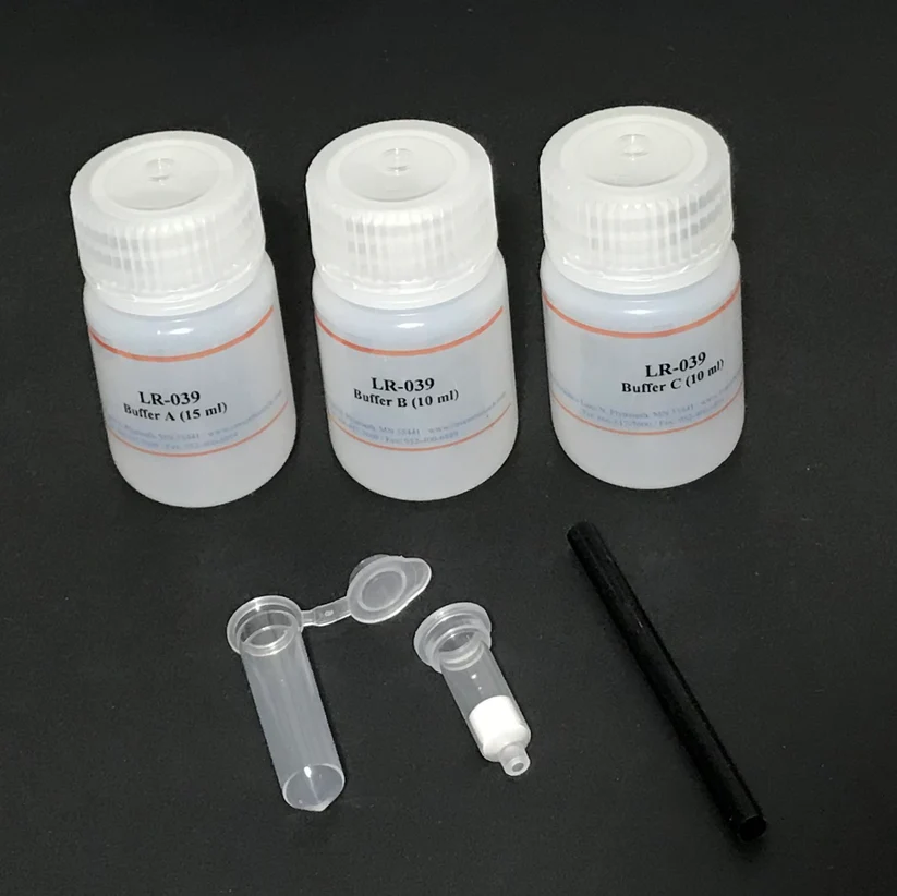
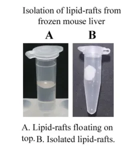
Lipid rafts are small membrane domains containing a high level of cholesterol and sphingolipids.  Lipid rafts are found in the plasma membrane (PM) and internal organellar membranes such as mitochondria-associated membrane (MAMs) and endoplasmic reticulum. They have been implicated in numerous cellular processes including but not limited to signal transduction, membrane trafficking, and protein sorting. Lipid-modified proteins and some transmembrane proteins are concentrated in the rafts while other proteins are excluded. Lipid rafts are also found to be associated with Na+/K+ ATPase on the plasma membrane. Lipid rafts are isolated by sucrose-gradient or OptiPrep gradient traditionally using ultracentrifugation that requires a large amount of starting material. The protocol is tedious and time-consuming. This kit was developed using our proprietary spin-column-based technologies, offering a rapid and easy way to isolate lipid rafts. Total membrane fraction (include PM and organelle membranes) is first isolated and treated with a non-ionic detergent containing buffer, followed by isolation of detergent-resistant fraction by flotation centrifugation using just a tabletop microcentrifuge. Highly enriched total lipid rafts can be obtained from cultured cells/tissues in less than 90 min without using density gradient and ultracentrifugation.


*For isolation of plasma membrane-derived lipid rafts, please refer to MinuteTM Plasma Membrane-Derived Lipid Raft Isolation Kit under  Cat #: LR-042

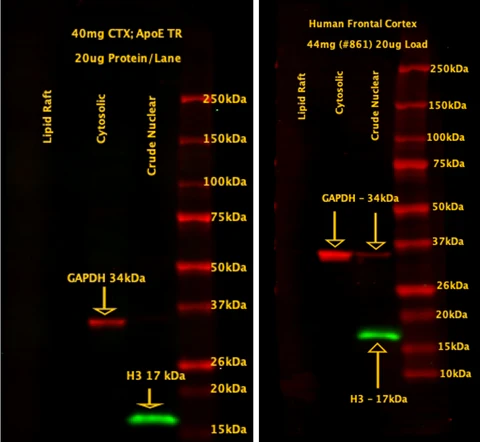
Evaluation of Cross-Contamination of Isolated Lipid Rafts

Evaluation of Cross-Contamination of Isolated Lipid Rafts

Lipid rafts were isolated from mouse (TR, left) and human (right) cortex that were probed with H3 and GAPDH. No presence of either in the lipid raft fraction. Human crude nuclear extract has some GAPDH, likely from RBCs. Data: Courtesy of Dr. Max Thorwald, University of Southern California.

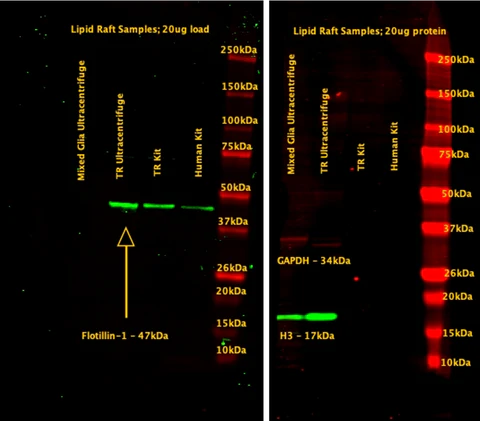
Detection of Lipid Raft Marker in Isolated Lipid Rafts

Lipid raft fractions obtained from isolation kit for mouse (TR, left) and human cortex (right) were probed with Flotillin-1. Lipid raft fractions are positive for Flotillin-1.  Data: Courtesy of Dr. Max Thorwald, University of Southern California.

Comparison of Lipid Raft Extractions kit with Ultracentrifugation

Samples were probed with Flotillin-1 on the left. All lipid raft samples probed for H3 and GAPDH samples on the right. The isolation kit yields enriched lipid raft fractions and no visible presence of H3 or GAPDH. Data: Courtesy of Dr. Max Thorwald, University of Southern California.

*TR=transgenic mouse cortex; Human=Human Cortex


Kit includes:

Items

Quantity

Buffer A

15 ml

Buffer B

10 ml

Buffer C10 ml

Filter Cartridges

20 units

Collection Tubes (2 ml)

20 units

Plastic Rod

2

 References (11)

1. Wang, M., Liu, J., Tu, Y., Zhao, Z., Qu, J., Chen, K., … & Wu, C. (2020). RSU-1 interaction with prohibitin-2 links cell-extracellular matrix detachment to down-regulation of extracellular signal-regulated kinase signalling. Journal of Biological Chemistry, jbc-RA120.

2.Thorwald, M. A., Cacciottolo, M., Forman, H. J., Morgan, T. E., & Finch, C. E. (2021). Reductions in ApoE and GPx4 highlight the Alzheimer’s disease lipid raft vulnerability. Alzheimer’s & Dementia, 17, e054511.https://doi.org/10.1002/alz.054511

3. Thorwald, M. A., Silva, J., Head, E., & Finch, C. E. (2022). Amyloid futures in the expanding pathology of brain aging and dementia. Alzheimer’s & Dementia.

4. Thorwald, M. A., & Finch, C. E. (2022). Alzheimer brains: increased soluble amyloid peptides, but decreased lipid rafts. Alzheimer’s & Dementia18, e063802.

5.  Edwards-Hicks, J., Apostolova, P., Buescher, J. M., Maib, H., Stanczak, M. A., Corrado, M., … & Pearce, E. L. (2023). Phosphoinositide acyl chain saturation drives CD8+ effector T cell signaling and function. Nature Immunology, 1-15.

6.  Thorwald, M., Godoy-Lugo, J. A., Silva, J., Head, E., O’Day, P. A., Morgan, T. E., … & Finch, C. E. (2023). Alzheimer’s Disease associations of ferritin and glutathione with oxidative damage and neuronal loss. bioRxiv, 2023-03.

7.  Thorwald, M. A., Godoy‐Lugo, J. A., Forman, H. J., & Finch, C. E. (2023). ApoE4 is associated with higher lipid peroxidation but not protein nitration in AD brains. Alzheimer’s & Dementia19, e079300.

8.  Lei, Y., Yang, H., Xu, X., Tan, Q., Xiang, Y., Tan, T., … & Xie, P. (2023). Inducing the Localization of HSP70 to Lipid Rafts by Borna Disease Virus 1 Promotes Its Invasion and Host Cells Apoptosis.

9. McGill, C. J., Christensen, A., Qian, W., Thorwald, M. A., Lugo, J. G., Namvari, S., … & Pike, C. J. (2024). Protection against APOE4-associated aging phenotypes with the longevity-promoting intervention 17α-estradiol in male mice. bioRxiv, 2024-03.

10.  Rossi, M., Banskota, N., Shin, C. H., Anerillas, C., Tsitsipatis, D., Yang, J. H., … & Gorospe, M. (2024). Increased PTCHD4 expression via m6A modification of PTCHD4 mRNA promotes senescent cell survival. Nucleic Acids Research, gkae322.

11.  Chaudhary, N., Kasiewicz, L. N., Newby, A. N., Arral, M. L., Yerneni, S. S., Melamed, J. R., … & Whitehead, K. A. (2024). Amine headgroups in ionizable lipids drive immune responses to lipid nanoparticles by binding to the receptors TLR4 and CD1d. Nature Biomedical Engineering, 1-16.

Minute™ Plasma Membrane-Derived Lipid Raft Isolation Kit

Home / Articles posted byadmin

Minute™ Plasma Membrane-Derived Lipid Raft Isolation Kit (20 preps)

Cat# : LR-042

Description

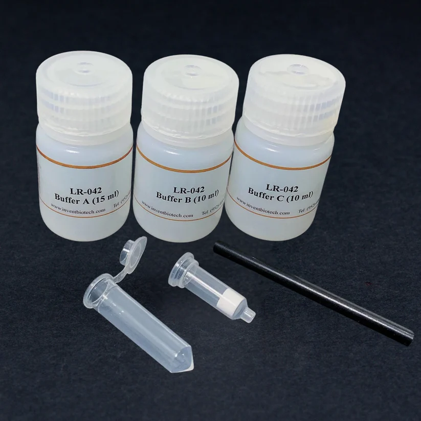
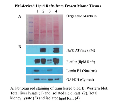
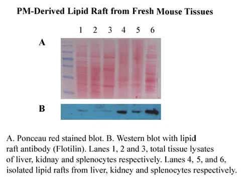
Lipid rafts are small membrane domains containing a high level of cholesterol and sphingolipids. Lipid rafts have been found in the plasma membrane (PM) and internal organellar membranes such as mitochondria-associated membrane (MAMs) and endoplasmic reticulum. Lipid rafts are implicated in numerous cellular processes such as signal transduction, membrane trafficking, and protein sorting.  Lipid-modified proteins and some transmembrane proteins are concentrated in the rafts while other proteins are excluded. Lipid rafts are also found to be associated with Na+/K+ ATPase on PM. Traditional methods for lipid raft isolation involve isolation of detergent-resistant membrane subdomain from total membranous structures, which does not distinguish plasma membrane-derived and/or organelle-derived lipid rafts. Using the patented spin-column-based technologies, we have developed this kit specifically for the isolation of plasma membrane-derived lipid rafts. Larger plasma membrane vesicles are first isolated and treated with a non-ionic detergent containing buffer followed by isolation of detergent-resistant fraction by flotation centrifugation using a tabletop microcentrifuge. Highly enriched plasma membrane-derived lipid rafts can be obtained in about 1 hour without using a traditional homogenizer and ultracentrifugation.

*For total lipid raft isolation, please refer to MinuteTM Total Lipid Raft Isolation Kit under Cat # LR-039.

Kit Components:

Items

Quantity

Buffer A

15 ml

Buffer B

10 ml

Buffer C

10 ml

Plastic Rods

2 units

Filter Cartridge with Collection Tubes

20 units

 References (13)
  1. Bu, Y., Teng, Q., Feng, D., Sun, L., Xue, J., & Zhang, G. (2021). YLMY Tyrosine Residue within the Cytoplasmic Tail of Newcastle Disease Virus Fusion Protein Regulates Its Surface Expression to Modulate Viral Budding and Pathogenicity. Microbiology Spectrum9(3), e02173-21.
  2. Rashkovan, M., Albero, R., Gianni, F., Perez-Duran, P., Miller, H. I., Mackey, A. L., … & Ferrando, A. A. (2021). Intracellular cholesterol pools regulate oncogenic signaling and epigenetic circuitries in Early T-cell Precursor Acute Lymphoblastic Leukemia. Cancer discovery.
  3. Jiang, C., Lin, Y., Shan, H., Xia, W., Pan, C., Wang, N., … & Yu, X. (2022). miR-146a Protects against Staphylococcus aureus-Induced Osteomyelitis by Regulating Inflammation and Osteogenesis. ACS Infectious Diseases.
  4. Fiore, D., Proto, M. C., Franceschelli, S., Pascale, M., Bifulco, M., & Gazzerro, P. (2022). In Vitro Evidence of Statins’ Protective Role against COVID-19 Hallmarks. Biomedicines10(9), 2123.
  5. Lim, J. H., Ahmad, K., Chun, H. J., Hwang, Y. C., Qadri, A. F., Ali, S., … & Lee, E. J. (2022). IgLON4 Regulates Myogenesis via Promoting Cell Adhesion and Maintaining Myotube Orientation. Cells11(20), 3265.
  6. Sha, Y. L., Liu, Y., Yang, J. X., Wang, Y. Y., Gong, B. C., Jin, Y., … & Zhao, Q. (2022). B3GALT4 remodels the tumor microenvironment through GD2-mediated lipid raft formation and the c-met/AKT/mTOR/IRF-1 axis in neuroblastoma. Journal of Experimental & Clinical Cancer Research41(1), 1-21.
  7. Gao, S. S., Shi, R., Sun, J., Tang, Y., Zheng, Z., Li, J. F., … & Li, C. (2022). GPI-Anchored Ligand-BioID2-Tagging System Identifies Galectin-1 Mediating Zika Virus Entry. iScience, 105481.
  8. Li, B., Ding, Z., Calbay, O., Li, Y., Li, T., Jin, L., & Huang, S. (2022). FAP is critical for ovarian cancer cell survival by sustaining NF-κB activation through recruitment of PRKDC in lipid rafts. Cancer Gene Therapy, 1-14.
  9. Oh, S., Jo, S., Bajzikova, M., Kim, H. S., Dao, T. T., Rohlena, J., … & Park, S. (2023). Non-bioenergetic roles of mitochondrial GPD2 promote tumor progression. Theranostics13(2), 438.
  10. Zhao, Q., Su, H., Jiang, W., Luo, H., Pan, L., Liu, Y., … & Tan, B. (2023). IGF-1 Combined with OPN Promotes Neuronal Axon Growth in Vitro Through the IGF-1R/Akt/mTOR Signaling Pathway in Lipid Rafts. Neurochemical Research, 1-12.
  11. Mirza, I., Haloul, M., Hassan, C., Masrur, M., Mostafa, A., Bianco, F. M., … & Mahmoud, A. M. (2023). Adiposomes from Obese-Diabetic Individuals Promote Endothelial Dysfunction and Loss of Surface Caveolae. Cells12(20), 2453.
  12. Khalifa, B. A., Guijarro, A., Ravera, S., Bertola, N., Adorni, M. P., Papotti, B., … & Nencioni, A. (2023). Cyclic fasting bolsters cholesterol biosynthesis inhibitors’ anticancer activity. Nature Communications14(1), 6951.
  13. Cheng, M., Zhang, R., Li, J., Ma, W., Li, L., Jiang, N., … & Wu, Z. (2024). MβCD inhibits SFTSV entry by disrupting lipid raft structure of the host cells. Antiviral Research, 106004.

Minute™ Chloroplast Isolation Kit

Home / Articles posted byadmin

Minute™ Chloroplast Isolation Kit (50 Preps)

Cat# : CP-011

Description

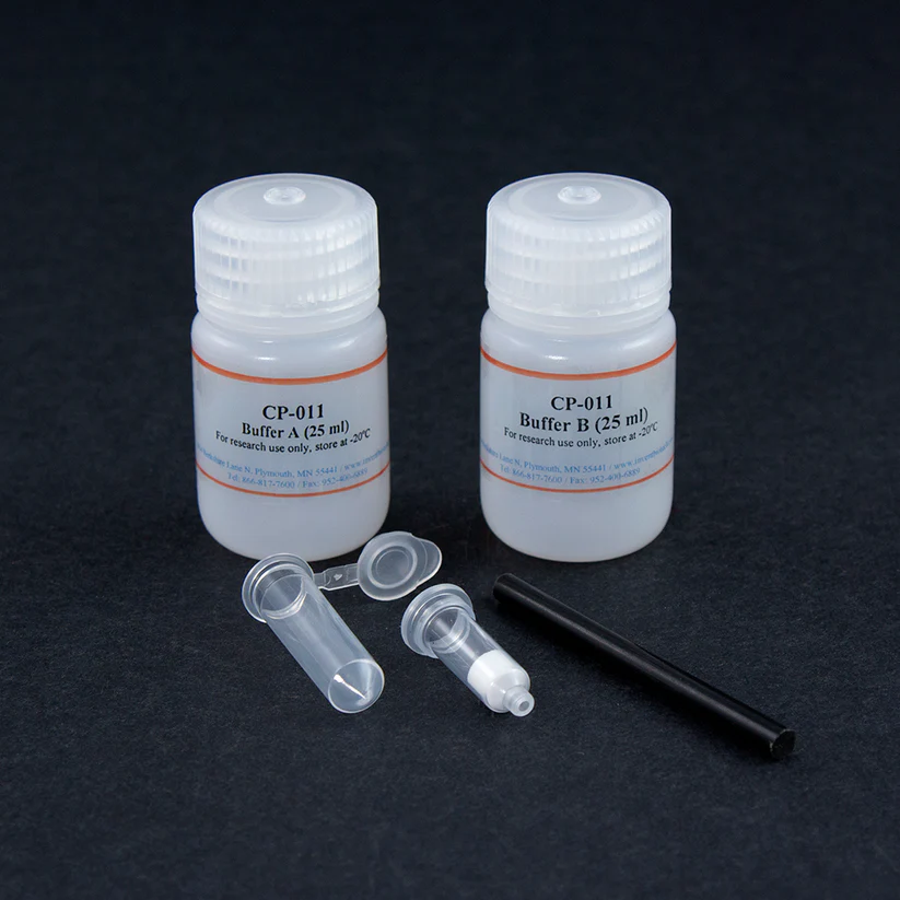
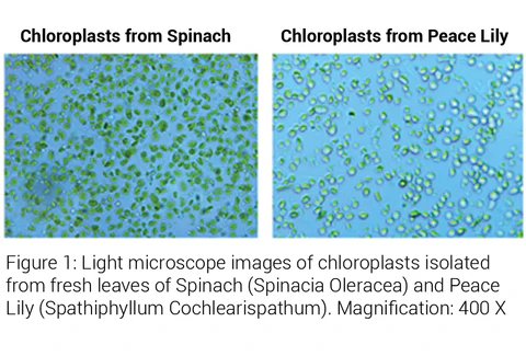
Invent Biotechnologies MinuteTM chloroplast isolation kit is composed of optimized chloroplast isolation buffers and filter cartridges with 2.0 ml collection tubes. The kit is designed to rapidly isolate intact chloroplasts from fresh plant tissues (leaves, seeds and soft stems etc.). Due to the use of filter cartridges with pre-defined pore size and thickness, intact chloroplasts can be isolated from 50-200 mg fresh plant tissues in less than 5 min. Unlike many other methods that require 1-10+ gram tissues for chloroplast isolation, this kit can quickly obtain 1 X 10⁶ to 1 X 10⁷ intact chloroplasts (>90% intact) from fresh plant leaves.

Kit includes: 

Items

Quantity

Buffer A (with 5 grams dissociation beads)

25 ml

 
Buffer B

25 ml

Protein Extraction Filter Cartridges

50 units

Collection Tubes with Caps

50 units

Plastic Rods

2 units


Minute™ Golgi Apparatus Enrichment Kit

Home / Articles posted byadmin

Minute™ Golgi Apparatus Enrichment Kit (20 Preps)

Cat# : GO-037

Description

The Golgi apparatus, also known as the Golgi complex or Golgi body, consists of a series of flattened stacked pouches called cisternae. This organelle plays a crucial role in eukaryotic cells by facilitating the transportation, modification, and packaging of proteins and lipids into vesicles for delivery to specific locations. The quantity and distribution of Golgi vary significantly across different cell and tissue types. Obtaining a highly enriched Golgi fraction is a crucial initial step in the study of its function and interactions with other organelles.

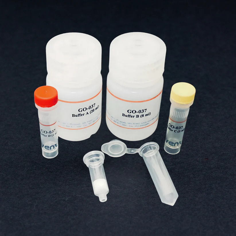
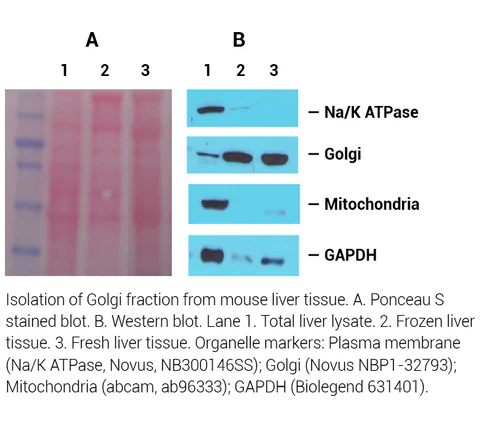
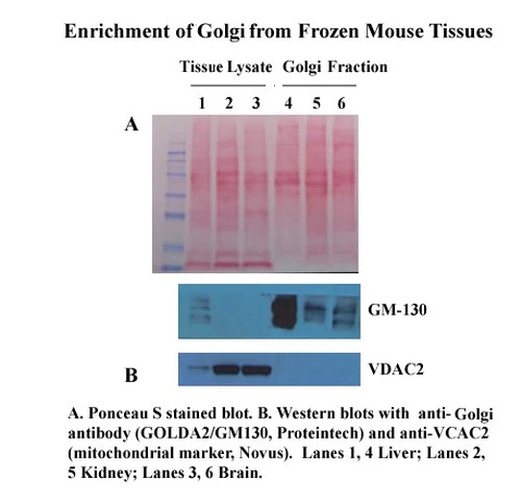
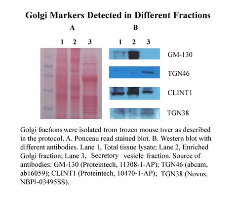
Traditional methods for isolating the Golgi apparatus rely on density gradient ultracentrifugation, which demands a substantial amount of starting material and can be laborious and time-consuming. In contrast, the Minute™ kit distinguishes itself from other Golgi isolation kits by employing patented spin-column-based technology. This approach is both straightforward and rapid, requiring only a small amount of starting material. With this kit, native Golgi can be preferentially enriched through precipitation, eliminating the need for a Dounce homogenizer and ultracentrifugation. It enables the isolation of two sub-Golgi fractions: the Golgi apparatus and secretory vesicles of the Golgi.

Kit includes:

Items

Quantity

Buffer A

20 ml

Buffer B

8 ml

Buffer C

2 ml

Buffer D2 ml

Plastic Rods

2 units

Filter Cartridge

20 units

Collection Tubes

20 units


Minute™ Cytosolic and Nuclear Extraction Kit for Frozen/Fresh Tissues

Home / Articles posted byadmin

Minute™ Cytosolic and Nuclear Extraction Kit for Frozen/Fresh Tissues (20 Preps)

Cat# : NT-032

Description

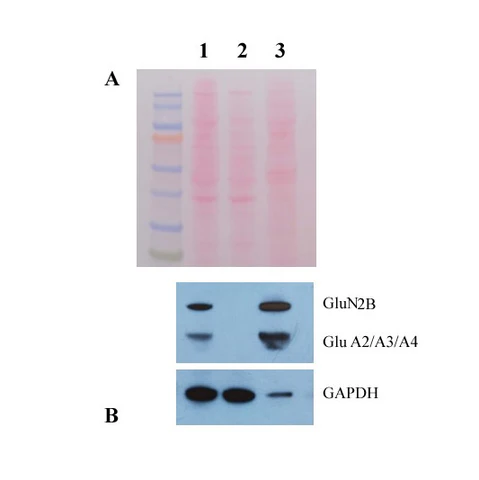
Preparation of cytosolic and nuclear fractions from cultured cells and tissues is common. Cell fractionation from cultured cells is relatively easy and straightforward. However, a clear separation of cytosolic and nuclear fractions from tissues, especially frozen tissues, is challenging. The cross-contamination from frozen tissues is always an issue because of the altered tissue cellular structure caused by the freeze and thaw cycle, improper homogenization, and the use of an inadequate extraction buffer. The majority of current commercial kits are designed to treat cultured cells and tissues similarly, not paying sufficient attention to the structural difference between the two. As a result, cross-contamination of cytosolic and nuclear fractions from fresh/frozen tissues remains a major problem (see references 1 and 2 below). This novel Cytosolic and Nuclear Extraction Kit is specially designed to address the issue by employing proprietary buffers and unique protocol to minimize the cross-contamination for fresh and frozen tissue samples.

Kit includes:

Items

Quantity

Buffer A

25 ml

Buffer B

10 ml

Buffer D

1.5 ml

Buffer N

1.5 ml

Pestle for 1.5 ml Tube

2 units

Protein Extraction Powder

2 grams


Minute™ ER Enrichment Kit

Home / Articles posted byadmin

Minute™ ER Enrichment Kit (20 Preps)

Cat# : ER-036

Description

The endoplasmic reticulum (ER) is a significant membranous structure that bridges the nuclear membrane and the plasma membrane, serving as a pivotal player in the exocytic pathway of protein trafficking in all eukaryotic cells. Proteins synthesized in the cytoplasm are directed to the ER, from where vesicles transport protein cargo to the Golgi apparatus, leading to subsequent fusion with the plasma membrane.

Traditional ER isolation methods rely on density gradient ultracentrifugation, a process demanding a substantial amount of starting material. These methods can be laborious, time-consuming, and often result in notable cross-contamination. Remarkably, all existing commercial kits for ER isolation are based on techniques developed in the 1970s. In a departure from these traditional approaches, the Minute™ ER enrichment kit stands out in the market by harnessing patented spin-column-based technology. This innovative method is not only straightforward and rapid but also requires only a small quantity of starting cultured cells or tissues.

This kit excels at differentially precipitating native ER, primarily rough ER, from cultured cells and tissues, all without the need for a Dounce homogenizer or ultracentrifugation. The entire protocol can be completed in approximately two hours.

Kit includes:

Items

Quantity

Buffer A

20 ml

Buffer B1 ml
Buffer C1 ml
Buffer D10 ml
Plastic Rods2 Units
Filter Cartridge20 Units
Collection Tubes20 Units


Quotation request