Minute™ Thylakoid Enrichment and Chloroplast Fractionation Kit

Home / Articles posted byadmin

Minute™ Thylakoid Enrichment and Chloroplast Fractionation Kit (20 preps)

Cat# : CF-055

Description

A chloroplast, delimited by a two-membrane envelope, is a type of plastid that serves as the site of photosynthesis. The photosynthetic function is based on developing an operational extensive internal membrane network, called the thylakoids, and on enzymatic processes in the chloroplast matrix, called the stroma. Thylakoid membrane is structurally different from the chloroplast envelope, and their biogenesis depends on biosynthetic and transporting activities specific to the chloroplast envelope. This kit is designed to fractionate chloroplasts into three parts (thylakoid membrane, envelope membrane, and stroma) using a specialized filter cartridge and differential centrifugation (without ultracentrifugation). Isolated thylakoids are in native forms and can be used for functional studies and the studies of subcellular localization of proteins using Western blotting/Mass spectrometry. The buffers in the kit are detergent and EDTA-free. The protocol can be completed in about one hour.

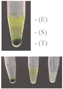
T: Left, S: Middle, E: Right

Figure 1. Fractionation of chloroplasts from spinach leaf into three fractions: Thylakoid (T), Stroma (S), and Envelope membranes (E).


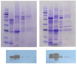
Figure 2. SDS-PAGE (Upper) and Western blot (Lower) of fractionated chloroplasts.   Lane 1. Total tissue lysate, 2. Thylakoid, 3. Stroma and 4. Envelope membranes. Thylakoid marker antibody was purchased from Agrisera (AS05084).

Kit Components:

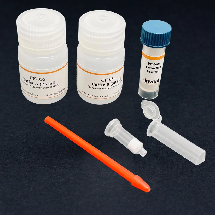
Buffer A25 ml
Buffer B30 ml
Filter Cartridge / 2.0 ml Collection Tube40 units
Pestles for 1.5 ml Eppendorf Microtubes 2 units
Protein Extraction Powder5g

Minute™ Detergent-Free Nuclear Matrix Protein Extraction Kit

Home / Articles posted byadmin

Minute™ Detergent-Free Nuclear Matrix Protein Extraction Kit (For Mammalian Cultured Cells/Tissues) (20 Preps)

Cat# : NM-033

Description

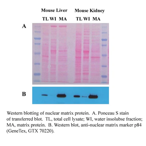
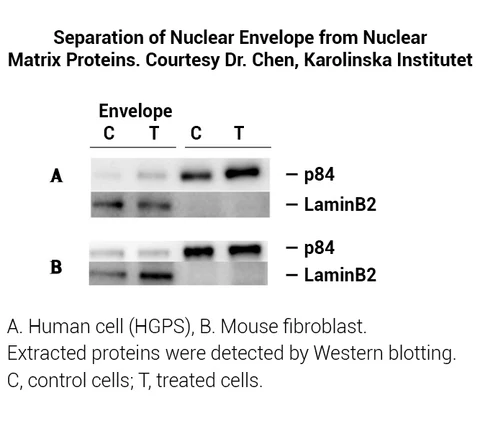
Nucleoplasm, also referred to as karyoplasm/nucleus sap, consists of chromosomes, nucleolus and nuclear matrix. Nucleoplasm plays a central role in genetic information flow and regulation of gene expression. Nuclear protein isolation has traditionally been accomplished by extraction protocol using detergent-containing buffers, which is effective in extracting total nuclear proteins but unable to separate membrane-bound proteins and water-soluble matrix proteins. The detergent-free nuclear matrix protein extraction kit is designed to separate cellular protein into three fractions: cytosolic, nuclear matrix and water-insoluble fractions (mainly envelope and nucleic acid-associated proteins) using a patented spin-column-based technology. The buffers used are detergent and EDTA-free. The whole protocol can be completed in less than 1 hour. The proteins extracted are in their native state and can be used in many downstream applications such as gel retardation, transcription factor analysis, apoptosis and protein trafficking studies.  

Kit includes:

Items

Quantity

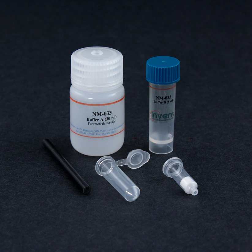
Buffer A

30 ml

Buffer B

5 ml

Protein Extraction Filter Cartridges

20 units

Plastic Rods

2 units

Collection Tubes

20 units

References (1)
  1. Luo, Q., Wu, X., Zhao, P., Nan, Y., Chang, W., Zhu, X., … & Liu, Z. (2021). OTUD1 Activates Caspase‐Independent and Caspase‐Dependent Apoptosis by Promoting AIF Nuclear Translocation and MCL1 Degradation. Advanced Science, 2002874.

Minute™ Plant ER Enrichment Kit

Home / Articles posted byadmin

Minute™ Plant ER Enrichment Kit (20 preps)

Cat# : PR-048

Description

Isolation and enrichment of plant endoplasmic reticulum (ER) are essential for elucidating ER-related cellular functions. The traditional method for ER isolation is tedious and time-consuming.  In animal cells, ER is closely associated with microtubules, and Golgi apparatus are clustered at microtubule organizing center. The ER is associated with actin microfilaments in plant cells, and no microtubule organizing center is found near the nuclei. Due to the structural characteristics of plan ER, it is much more challenging to isolate/enrich plant ER than animal samples. Minute™ plant ER enrichment kit was specially designed for plants. One to three-fold enrichment of ER fraction can be obtained from plant leave samples in about 1 hour. The enriched ER is essentially free from Golgi apparatus contamination. The enriched ER fraction can be used in plant protein trafficking studies.

Kit Components:

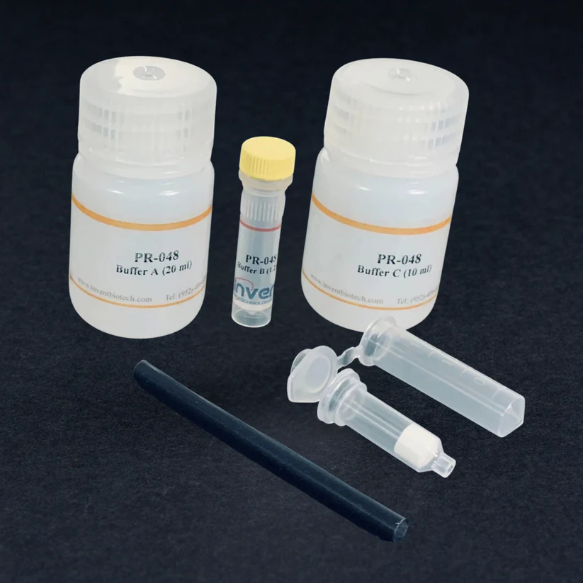
Buffer A 20 ml
Buffer B1.2 ml
Buffer C10 ml
Plastic Rods2
Filter Cartridge20
2 ml Collection Tibe20

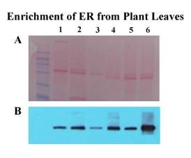
A. Ponceau S Stained Blot; B. Western blot with Anti-ER marker antibody (BIP2, Agrisera, Vannas, Sweden. AS 09481). Lanes 1&2: B. napus; Lanes 3&4: N. tabacum; Lanes 5&6: A. thaliana. Lanes 1, 3 & 5: Total Protein; Lanes 2, 4 & 6: ER fractions.

Minute™ High Fidelity ER Isolation Kit for Liver Tissue

Home / Articles posted byadmin

Minute™ High Fidelity ER Isolation Kit for Liver Tissue (20 Preps)

Cat# : ER-035

Description

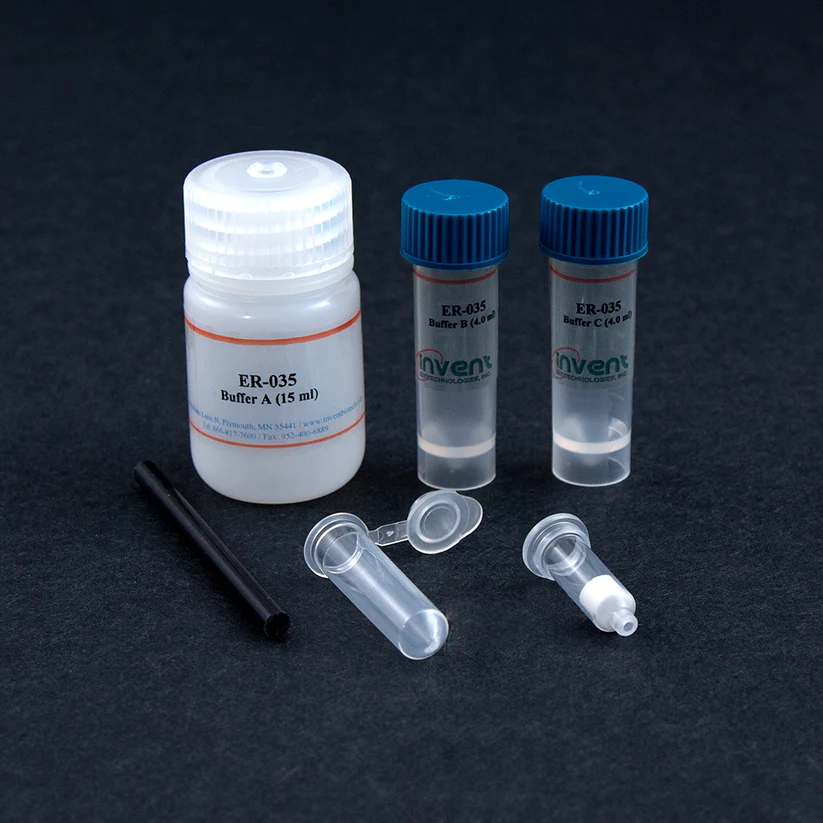
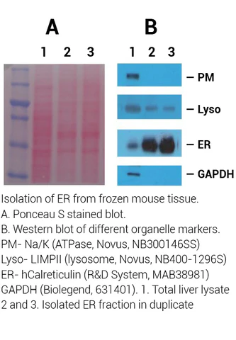
Endoplasmic reticulum (ER) is a major membranous structure that functionally connects nuclear membrane and plasma membrane. Liver is an ER-rich organ and has been used for isolation of ER for decades. Traditional methods for isolating ER are based on density-gradient ultracentrifugation. The protocol requires a large amount of starting material and the methods are tedious and time-consuming and contain significant cross-contamination. Currently, all commercial kits for ER isolation are based on methods developed in the 1970s. Unlike any other ER isolation kit on the market, this kit employs a patented spin-column-based technology that is simple and rapid and requires only a small amount of tissue. This kit can differentially precipitate native ER (mainly rough ER) from frozen liver tissues without using a Dounce homogenizer or ultracentrifugation. The whole protocol can be done in about 2 hours.

Kit includes:

Items

Quantity

Buffer A

20 ml

Buffer B

4 ml

Buffer C

4 ml

Plastic Rods

2 units

Filter Cartridge

20 units

Collection Tubes

20 units

Minute™ Yeast/Fungus Nuclear and Cytoplasmic Protein Extraction Kit

Home / Articles posted byadmin

Minute™ Yeast/Fungus Nuclear and Cytoplasmic Protein Extraction Kit (20 preps)

Cat# : YN-058

Description

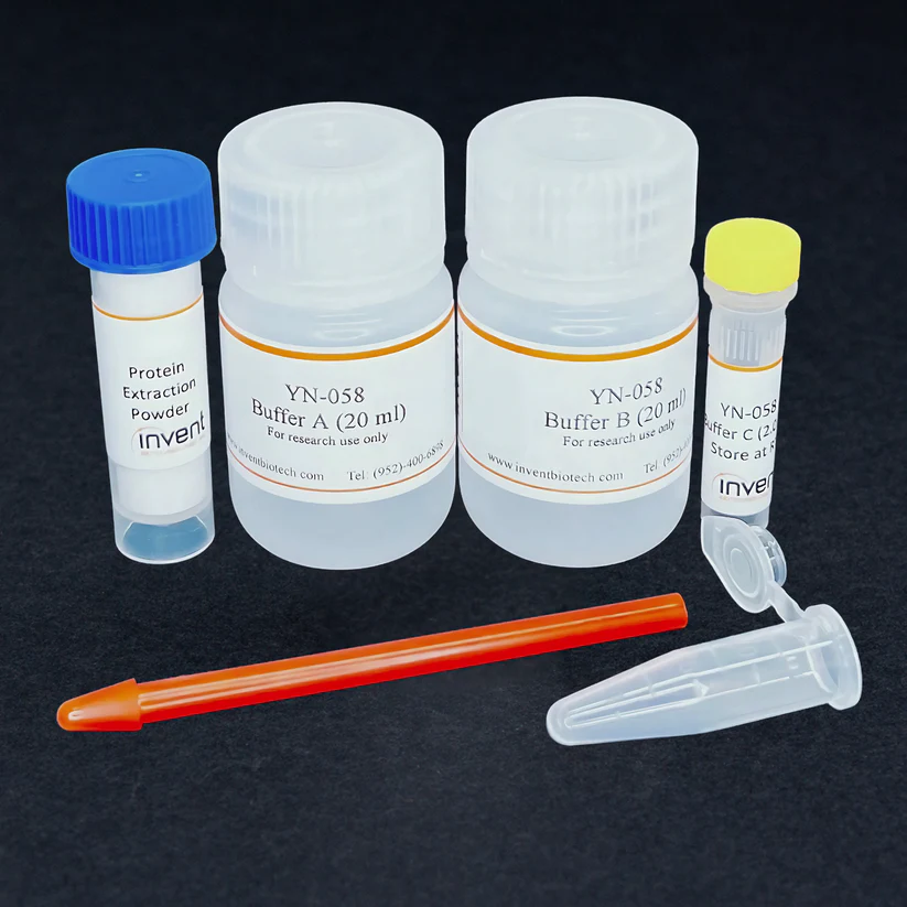
The Minute™ Yeast/Fungus Nuclear and Cytoplasmic Protein Extraction Kit offers a fast and reliable method to isolate both cytosolic and nuclear proteins from yeast and fungus. This easy-to-use kit allows you to separate these crucial cellular components in about 1h, streamlining your research workflow. The kit provides all the necessary buffers and disposable tools for an efficient protein extraction. Using this kit, you’ll have access to both cytosolic and intact nuclei that can be used for nuclear protein and nucleic acid extraction.

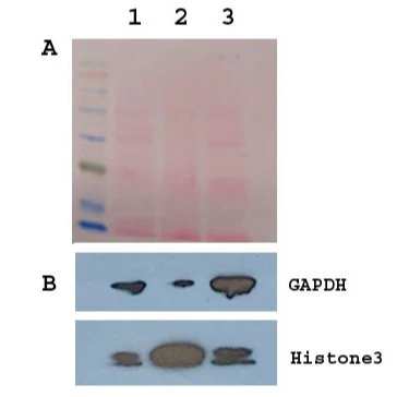
Fig. 1. Isolation of cytosolic and nuclear fractions from S. cerevisiae.

  1. Ponceau red stained protein profiles on nitrocellulose membrane.
  2. Western blotting using anti-GAPDH and Histone3.

Lane 1, total cell lysate. Lane 2, Nuclear fraction. Lane 3, cytosolic fraction.

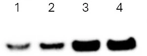
Fig. 2. Enhancement of Nuclear Signal by Western Blotting. Equal amounts of total cell lysate of S. cerevisiae (lane 1 and 2) and nuclear protein extract (lane 3 and 4) were separated on SDS-PAGE and probed with anti-histone3 antibody. Data provided by a third-party testing.

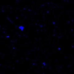
Fig. 3. Nuclei Isolated from S. cerevisiae. DAPI staining (Mag. 20 X)

Fig. 4. Nuclei Isolated from A. fumigatus Stained with DAPI. (Mag. 20 X).

(Courtesy of Sandeep Dartmouth School of Medicine.)

Kit includes:

Items

Quantity

Buffer A

20 ml

Buffer B

20 ml

Buffer C

2 ml

1.5 ml Microfuge Tubes

20 units

Pestles for 1.5 ml Tubes

2 units

Protein Extraction Powder

5 grams


Minute™ Yeast Mitochondria Enrichment Kit

Home / Articles posted byadmin

Minute™ Yeast Mitochondria Enrichment Kit (50 Preps)

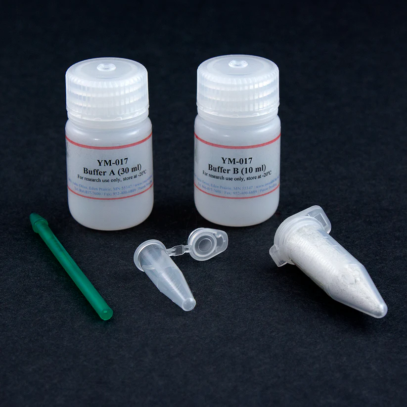
Cat# : YM-017

Description

Traditional protocol for yeast mitochondria isolation/extraction involves a range of centrifugation-based sub-cellular fractionation procedures. Typically, the techniques include spheroplast preparation, glass-bead lysis using a homogenization instrument, differential centrifugation and several density gradient procedures using a variety of gradient media with ultracentrifugation. The procedures are very tedious and time-consuming. We feature a simple and efficient protocol for yeast mitochondria enrichment. The procedure is gentle and instrument free. Native mitochondrial proteins can be isolated from yeast in about one hour without ultracentrifugation. This kit contains optimized detergent-free protein extraction buffers. The protein yield is in the range of 150-250 μg/sample. The materials provided are sufficient for 50 extractions.

Kit includes:

Items

Quantity

Buffer A

30 ml

Buffer B

10 ml

Protein Extraction Powder

5 grams

1.5 ml Microfuge Tube

50 units

Pestles

4 units


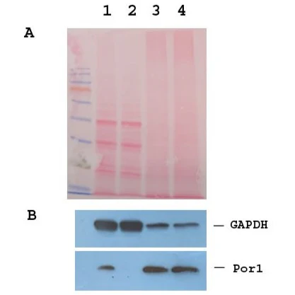
Enrichment of Mitochondria detected by Western Blotting.  Ponceau red stained blot (A) and Western blotting (B). Lane 1, Total yeast cell lysate; Lane 2, Cytosolic fraction; Lane 3 and 4, Mitochondrial fraction from two separated Experiments. Por1 is a mitochondrial marker antibody (Thermofisher, 16G9E6BC4).

References (1)
  1. Yamashita, K., Miyazaki, T., Fukuda, Y., Mitsuyama, J., Saijo, T., Shimamura, S., … & Kohno, S. (2019). The novel arylamidine T-2307 selectively disrupts yeast mitochondrial function by inhibiting respiratory chain complexes. Antimicrobial Agents and Chemotherapy, AAC-00374.

Minute™ Plant Microsomal Membrane Extraction Kit

Home / Articles posted byadmin

Minute™ Plant Microsomal Membrane Extraction Kit (50 Preps)

Cat# : MM-018

Description

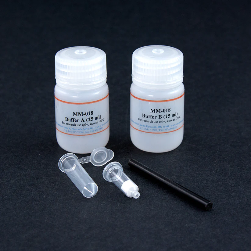
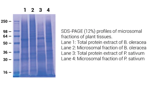
Isolation of microsomal membranes from plant tissues is a common procedure. The microsomal fraction of plant cell lysate is the focus of interest in many plant research projects and is believed to be enriched for plasma membranes, endoplasmic reticulum, Golgi apparatus, vacuolar membranes, and other components of a membrane system. Traditional methods for microsomal fraction isolation involve differential pelleting, where centrifugation steps are required for membrane fractions. Microsomal fraction isolation usually requires a large amount of starting material and has tedious ultracentrifugation steps. MM-018 offers a simple, fast, and user-friendly approach for microsomal membrane extraction with a small amount of starting material. During the procedures, water-soluble cytosolic proteins are removed, and water-insoluble microsomal fraction, especially plasma membrane fraction, is extracted with optimized buffers in a tabletop microcentrifuge. Native microsomal proteins can be isolated from plant tissue in about one hour without ultracentrifugation. The protein yield is in the range of 100-200 µg/sample.

Kit includes:

Items

Quantity

Buffer A

25 ml

Buffer B

15 ml

Filter Cartridges

50 units

2.0 ml Collection Tubes with Caps

50 units

Plastic Rods

2 units


References (4)
  1. Kim, S., Choi, Y., Kwon, C., & Yun, H. S. (2018). Endoplasmic reticulum stress‐induced accumulation of VAMP721/722 requires CALRETICULIN 1 and CALRETICULIN 2 in Arabidopsis. Journal of integrative plant biology.
  2. Yang, Z., Yang, J., Wang, Y., Wang, F., Mao, W., He, Q., … & Mao, C. (2020). PROTEIN PHOSPOHATASE 95 Regulates Phosphate Homeostasis by Affecting Phosphate Transporter Trafficking in Rice. The Plant Cel
  3. Yu, F., Cao, X., Liu, G., Wang, Q., Xia, R., Zhang, X., & Xie, Q. (2020). ESCRT-I Component VPS23A Is Targeted by E3 Ubiquitin Ligase XBAT35 for Proteasome-Mediated Degradation in Modulating ABA Signaling. Molecular Plant13(11), 1556-1569.
  4. Kato, T., Morita, R., Ootsuka, S., Wakabayashi, Y., Aoki, N., & Horibata, A. Evaluation of alleles at OsAGPS2, OsAGPL2, and OsSUT1 related to grain filling in rice in a common genetic background. Crop Science.
  5. Hou, L. Y., Lehmann, M., & Geigenberger, P. (2021). Thioredoxins o1 and h2 Show Different Subcellular Localizations and Redox-Active Functions, but Cooperatively Affect NADPH Redox Poise and Photosynthetic Performance in Fluctuating Light.
  6. Chiu, C. Y., Tsai, C. D., Wang, J. Y., Tsai, M. H., Kanno, S., Lung, H. F., & Liu, T. Y. (2024). Phosphate Starvation-Induced CORNICHON HOMOLOG 5 as Endoplasmic Reticulum Cargo Receptor for PHT1 Transporters in Arabidopsis. bioRxiv, 2024-06.

Minute™ Adipose Tissue Fractionation Kit

Home / Articles posted byadmin

Minute™ Adipose Tissue Fractionation Kit (20 Preps)

Cat# : AF-023

Description

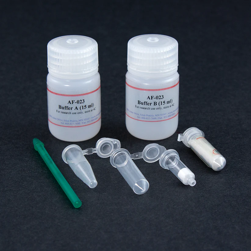
Total cellular proteins account for less than 2% of adipose tissue. The white adipose tissue, in particular, has been recognized as an essential endocrine and inflammation organ in addition to its energy storage function. Fractionation and analysis of proteins from adipose tissue are critical for understanding many physiological/pathological conditions. However, fractionation of adipose tissue is challenging due to its high lipid and low protein contents. The water-oil emulsion present in biological samples is notoriously difficult to separate. Still, we have developed a cutting-edge technology to deal with this issue: a porous filter with unique surface properties and predefined pore size coupling with a specially formulated detergent-free fractionation buffer. This is the key to fractionating adipose tissue into two: water-soluble protein fraction and water-insoluble fraction. The buffers used in this kit are free of primary amine, detergent, and reducing agents. Isolated proteins are compatible with all downstream applications, including TMT labeling, enzyme digestion, MS analysis, and other applications.

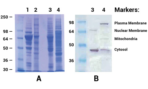
A. SDS-PAGE (10%) profiles of fractionated adipose tissues. Lane 1, a water-soluble fraction of porcine WAT; Lane 2, a water-insoluble fraction of porcine WAT; Lane 3, a water-soluble fraction of rat WAT; Lane 4, a water-insoluble fraction of rat WAT.

B. Western blottings of fractionated proteins from rat WAT; Lane 3. water-soluble fraction; Lane 4. Water-insoluble fraction. Proteins were separated in 10% SDS-PAGE and probed with the following cellular protein marker antibodies: Anti-Na/K ATPase alpha1, a plasma membrane marker (Upstate, clone 464.6), anti-lamin B1, a nuclear envelope marker (ab16048, Abcam Cambridge, MA), anti-ubiquinol-cytochrome C reductase core protein (Abcam, ab 96333) and GAPDH, a cytosolic marker (Sigma). The specific protein bands were visualized by a substrate Opti-4CN (Bio-RAD). 

Kit includes:

Items

Quantity

Buffer A

15 ml

Buffer B

15 ml

1.5 ml Microfuge Tubes

20 units

Pestles for 1.5 ml Tubes

2 units

Filter Cartridge with Collection Tubes

20 units

Protein Extraction Powder

2 grams


References (3)
  1. Markan, K. R., Naber, M. C., Small, S. M., Peltekian, L., Kessler, R. L., & Potthoff, M. J. (2017). FGF21 resistance is not mediated by downregulation of beta-klotho expression in white adipose tissue. Molecular Metabolism. http://dx.doi.org/10.1016/j.molmet.2017.03.009
  2. Liu, X., Tong, W., Zhao, X., Zhang, H., Tang, Y., & Deng, X. Chinese herb extract improves liver steatosis by promoting the expression of high molecular weight adiponectin in NAFLD rats. Molecular Medicine Reports.
  3. Subbaramaiah, K., Iyengar, N. M., Morrow, M., Elemento, O., Zhou, X. K., & Dannenberg, A. J. (2018). Prostaglandin E2down-regulates sirtuin 1 (SIRT1) leading to elevated levels of aromatase, providing insights into the obesity-breast cancer connection. Journal of Biological Chemistry, jbc-RA118.

Minute™ Mitochondria Isolation Kit for Muscle Tissues/Cultured Muscle Cells

Home / Articles posted byadmin

Minute™ Mitochondria Isolation Kit for Muscle Tissues/Cultured Muscle Cells (50 preps)

Cat# : MM-038

Description

Muscle cells have large number of mitochondria because they are continually used to move the body.  Another mitochondrion-rich organ is the heart where mitochondria make up about 40% of the heart cells. However, isolation of mitochondria from muscle tissues can be difficult due to that fact that interfibrillar mitochondria are hidden deep among muscle fibrils. Traditional methods usually require polytron tissue disintegrator and ultracentrifuge for purification with high density medium such as Percoll gradient. This kit was developed using our proprietary technologies, making mitochondria isolation from muscles simple, rapid and user friendly. Intact mitochondria can be isolated from fresh/frozen muscle tissues in about 1h with higher yield.

Kit includes:

Items

Quantity

Buffer A

25 ml

Buffer B

10 ml

Filter Cartridges

50 units

Collection tubes with cap (2 ml)

50 units

Plastic rods

2 units

Tissue dissociation beads

5 grams


References (5)
  1. Li, S., Dong, S., Xu, Q., Shi, B., Li, L., Zhang, W., … & Zhong, M. (2022). Sleeve Gastrectomy-Induced AMPK Activation Attenuates Diabetic Cardiomyopathy by Maintaining Mitochondrial Homeostasis via NR4A1 Suppression in Rats. Frontiers in Physiology13, 837798.
  2. Peng, L., Li, L., Fan, H., Lin, F., Liang, X., Zhu, Y., … & Sachinidis, A. (2023). Unmusking of Protein Phosphatase 2 Regulatory Subunit B as a crucial factor in the development and progression of dilated cardiomyopathy.
  3. Radajewska, A., Szyller, J., Krzywonos-Zawadzka, A., Olejnik, A., Sawicki, G., & Bil-Lula, I. (2023). Mitoquinone Alleviates Donation after Cardiac Death Kidney Injury during Hypothermic Machine Perfusion in Rat Model. International Journal of Molecular Sciences24(19), 14772.
  4. Seo, Y. K., Yoo, Y., Yeon, M., Kim, W. K., Shin, H. B., Lee, S. M., … & Ro, H. (2023). Age-dependent loss of Crls1 causes myopathy and skeletal muscle regeneration failure.
  5. Lin, F., Liang, X., Meng, Y., Zhu, Y., Li, C., Zhou, X., … & Peng, L. (2024). Unmasking Protein Phosphatase 2A Regulatory Subunit B as a Crucial Factor in the Progression of Dilated Cardiomyopathy. Biomedicines12(8), 1887.

Minute™ Synaptosome Isolation Kit

Home / Articles posted byadmin

Minute™ Synaptosome Isolation Kit (50 preps)

Cat# : SY-052

Description

The ability to isolate synaptosomes from neuronal tissues/cultured cells is essential for understanding the mechanisms of neurological diseases. Contained in synaptosomes are synaptic vesicles with diameters of 80-200 nm that play an important role in signal transmission. Traditionally, synaptosomes are isolated by density ultracentrifugation. The procedure is relatively tedious and time-consuming. A larger amount of starting material is usually required. MinuteTM synaptosome isolation kit provides a simple and rapid method for isolating synaptosomes from fresh/frozen neuronal tissues/cultured cells. The protocol can be completed in less than 1 h without using Dounce homogenizer and ultracentrifugation. The amount of starting material required (10-50 mg tissue) is only a fraction of that required by the traditional method. The buffers used are detergent-free and synaptosome associated proteins are isolated in native form.

Fig.1. Isolation of Synaptosomes from Fresh Mouse Cortex.

A, Ponceau S-stained blot. B, Western blot. Lane 1. Total tissue lysate;

Lane 2. Cytosolic fraction; Lane 3. Synaptosome fraction. Synaptosome

marker antibodies (GluN2B and Glu A2/A3/A4) are from Cell Signaling.


References (4)
  1.  Gu, J., Ke, P., Guo, H., Liu, J., Liu, Y., Tian, X., … & Xiao, F. (2023). KCTD13-mediated ubiquitination and degradation of GluN1 regulates excitatory synaptic transmission and seizure susceptibility. Cell Death & Differentiation, 1-16.
  2. Ke, P., Gu, J., Liu, J., Liu, Y., Tian, X., Ma, Y., … & Xiao, F. (2023). Syntabulin regulates neuronal excitation/inhibition balance and epileptic seizures by transporting syntaxin 1B. Cell Death Discovery9(1), 187.
  3. Han, X., Huang, S., Zhuang, Z., Zhang, X., Xie, M., Lou, N., … & Chen, S. (2023). Phosphatidate phosphatase Lipin1 involves in diabetic encephalopathy pathogenesis via regulating synaptic mitochondrial dynamics. Redox Biology, 102996.
  4. Zong R, Zhang X, Dong X, Liu G, Zhang J,Gao Y, Zhang Z, Ma Y, Gao H and Gamper N (2024) Genetic deletion of zinc transporter ZnT3 induces progressive cognitive deficits in mice by impairing dendritic spine plasticity and glucose metabolism. Front. Mol. Neurosci. 17:1375925. doi: 10.3389/fnmol.2024.1375925

Quotation request