Ultrasonic Cleaner BL-SB-5200DTD
Minute™ Single Nucleus Isolation Kit for Neuronal Tissues/Cells
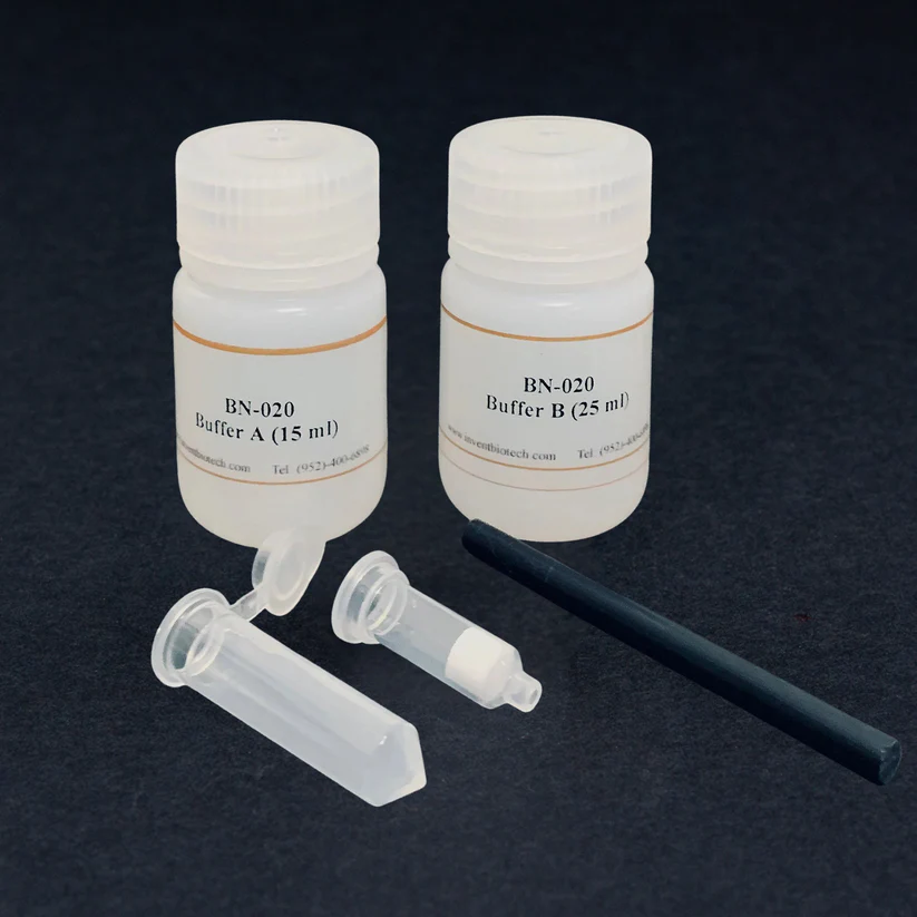
Minute™ Single Nucleus Isolation Kit for Neuronal Tissues/Cells (20 preps)
Cat #: BN-020
Description
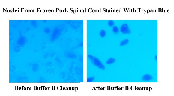
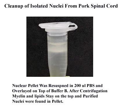
Isolated nuclei are widely used for various experiments such as FACS, single nucleus analysis (such as RNA-seq and ATAC-seq), immunofluorescence staining, cell cycle, and apoptosis research. Single-cell RNA-seq is a powerful technology for studying the complex cellular composition of tissues. However, Neurons are highly interconnected, and it is challenging to obtain single cells from neuronal tissues such as the brain and spinal cord. It is even more difficult to isolate intact cells from frozen neuronal tissues. Due to these limitations, single-cell RNA-seq is being substituted by single-nucleus-seq. The traditional method for single nucleus isolation from neuronal tissue is relatively tedious and time-consuming, and the yield is usually low because it is difficult to get rid of contaminating myelin and other cellular components. This kit is designed to address the issues with a simple, rapid, and effective protocol. The highly purified single nucleus can be obtained in about 30 min. Compared to the traditional method, the kit requires much less starting material and can handle an extensive range of sample sizes (1-25 mg). The buffers contain a proprietary mix of detergents for efficient cell lysis. If the presence of detergent is not desirable, a detergent-free nuclei isolation kit is also available under cat# NI-024.
For storage and transportation of isolated single nuclei, the following buffer is recommended:
WA-014: Minute™ Anti-Clumping Nuclei Storage Buffer
NOTE: We also offer the following single nucleus isolation kits with a much cleaner background if the presence of detergent is not a concern:
SN-047: Minute™ Single Nucleus Isolation Kit for Tissues/Cells
BN-020: Minute™ Single Nucleus Isolation Kit for Neuronal Tissues/Cells
AN-029: Minute™ Nuclei and Cytosol Isolation Kit for Adipose Tissues/Cultured Adipocytes
Kit includes:
Items | Quantity |
Buffer A | 15 ml |
Buffer B | 30 ml |
Filter Cartridges | 20 units |
Collection Tubes | 20 units |
Pestles | 2 units |
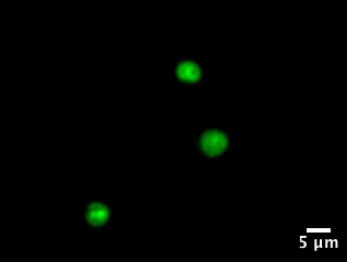
Nuclei Isolated from zebrafish brain tissue Using NI-024 and stained with DAPI (False-coloured in green) (Courtesy Sema Elif Eski, Singh Lab, Université Libre de Bruxelles).
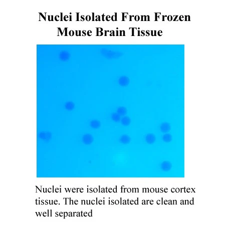
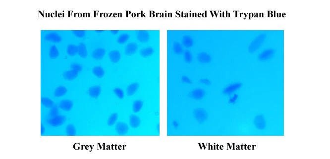
Kit includes:
Items | Quantity |
Buffer A | 15 ml |
Buffer B | 25 ml |
Pestles for 1.5 ml Tubes | 2 units |
Filter Cartridge with Collection Tubes | 20 units |
Minute™ Chloroplast Isolation kit for Mucilaginous Plants
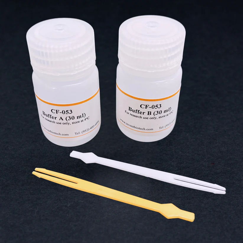
Minute™ Chloroplast Isolation kit for Mucilaginous Plants (20 preps)
Cat# : CF-053
Description
Many crop species across the plant family contain high polysaccharide and polyphenols. The hydrocolloid mucilage is a complex mucopolysaccharide typically showing high viscosity when released in solution. Homogenate of high mucopolysaccharide plant samples usually imparts a slimy consistency that is most unwanted contaminants that interfere with cell fractionation and organelle purification, such as chloroplast and nuclear isolation. This kit provides a simple, rapid, and efficient method for isolating chloroplasts from viscous plant homogenate. The protocol takes about 30 min.
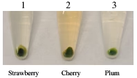
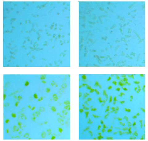
Figure 1. Chloroplasts isolated from leaves of indicated plant species.
Figure 2. Morphologies of chloroplasts isolated from leaves of indicated plants under light microscopy.
Kit Components:
| Buffer A | 30 ml |
| Buffer B | 30 ml |
| Micro Puncher | 2 |
Minute™ Water-soluble and Water-Insoluble Protein Fractionation Kit
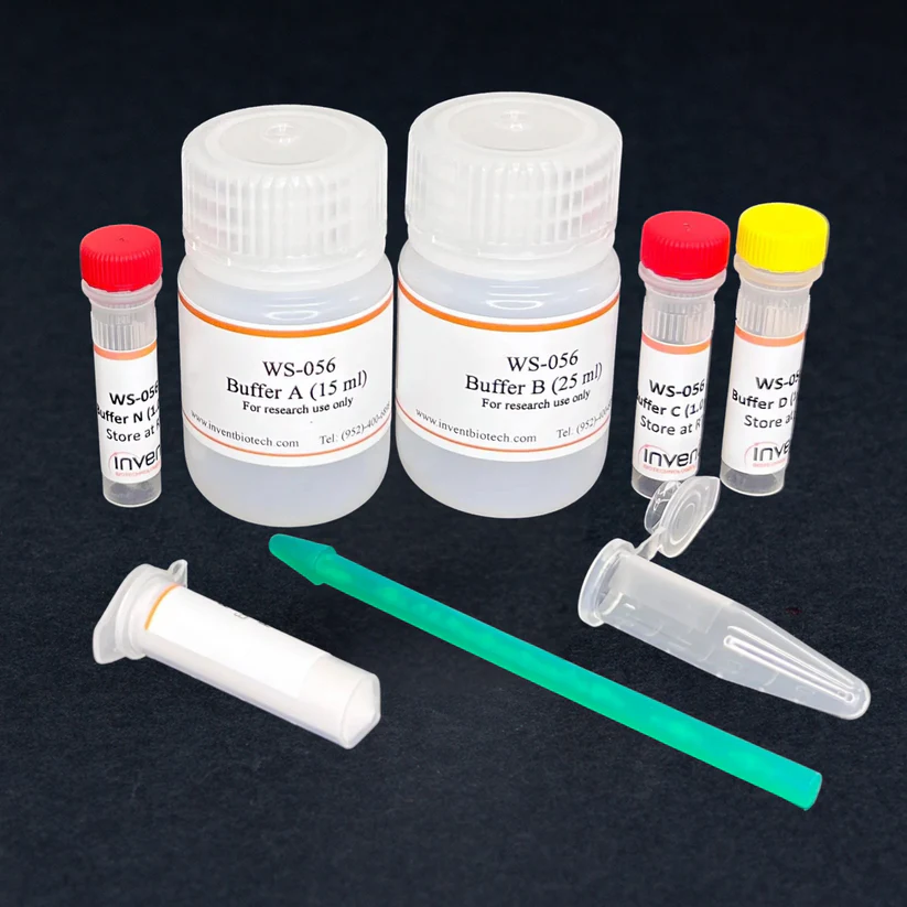
Minute™ Water-soluble and Water-Insoluble Protein Fractionation Kit (20 preps)
Cat# : WS-056
Description
Researchers studying protein localization and trafficking now have a convenient tool: the MinuteTM Water-soluble and Water-Insoluble Protein Fractionation Kit for cells/tissues. This unique product allows for the rapid and efficient separation of these two key fractions from cells/tissues, microorganisms and plant tissues, providing enriched and easily detectable protein samples for downstream applications like western blotting, ELISA and immunoprecipitation. Water soluble fraction contains mainly cytosolic proteins and water-insoluble fraction contains mainly insoluble membranous structures such as plasma membrane, nuclear membrane and organelles. The kit offers researchers a novel and simple approach for understanding protein function and movement within the cellular landscape. If a protein target is known to be localized in water-soluble or water-insoluble fraction, the kit will facilitate detection, isolation and purification of the target protein by enrich corresponding fractions.
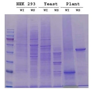
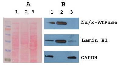
Fig. 1. Protein profiles of water-insoluble (WI) and water-soluble (WS) fractions from HEK293 cells, yeast (S. cerevisiae) and plant (Broccoli leaf). Proteins were separated in 10% SDS-PAGE and stained with Coomassie blue.
Fig. 2. Western blotting of fractionated frozen mouse liver. Lanes 1, 2 and 3 are total liver lysate, water-insoluble and water-soluable fraction, respectively. A. Ponceau red stained blot.
B. Western blot with anti-Na/K-ATPase (plasma membrane marker), Lamin B1 (nuclear envelope marker) and GAPDH (cytosolic marker).
Kit includes:
Items | Quantity |
Buffer A | 15 ml |
Buffer B | 25 ml |
Buffer C | 1 ml |
Buffer D | 1 ml |
Buffer N | 1 ml |
1.5 ml Microfuge Tubes | 20 units |
Pestles for 1.5 ml Tubes | 2 units |
Protein Extraction Powder | 2 grams |
Minute™ Plant Lipid Raft Isolation Kit
Description
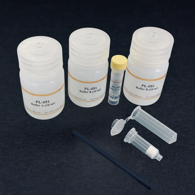
There are substantial evidences that support the existence of detergent-resistant membrane (DRM) subdomains in both animal and plant membrane systems, which is also referred to as lipid rafts. Lipid rafts are shown to have increased sphingolipid to protein ratio and higher cholesterol concentration. The lipid rafts are believed to play an important role in signal transduction and protein trafficking in eukaryotic cells. Traditional method for lipid raft isolation involves cold non-ionic detergent extraction followed by sucrose gradient ultracentrifugation. The protocol requires large amount of starting material (hundred grams amount) and specialized equipment in addition to lengthy protocol time. We have developed a rapid method for isolation of lipid rafts from plant tissues using only milligram amount of starting material and the protocol can be completed in about 1h without using ultracentrifugation.
Kit includes:
Buffer A | 10 ml |
Buffer B | 10 ml |
Buffer C | 1.2 ml |
Buffer D | 10 ml |
Plastic Rods | 2 units |
Filter Cartridge with Collection Tubes | 20 units |
Protein Extraction Powder | 2 grams |
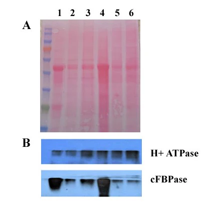
Western Blotting of Isolated Lipid Rafts. A. Ponceau red stained blot membrane. B. Western Blot. Samples: Leaves of Spinacia oleracea (1,2,3) and Blassica rapa (Lanes 4,5,6). Lanes 1 & 4, total tissue lysates. Lanes 2,3 & 4,5; isolated Lipid rafts. Rabbit anti H+ATPase (plasma membrane marker), and cFBPase (cytosolic marker) were purchased from Agrisera (Vannas, Sweden).
Reference (1)
1. Zhou, N., Li, X., Zheng, Z., Liu, J., Downie, J. A., & Xie, F. (2024). RinRK1 enhances NF receptors accumulation in nanodomain-like structures at root-hair tip. Nature Communications, 15(1), 1-16.
Minute™ Nuclear Proteasome Enrichment Kit
Description
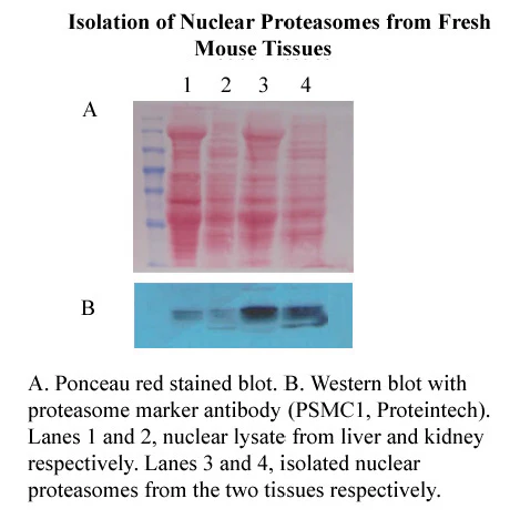
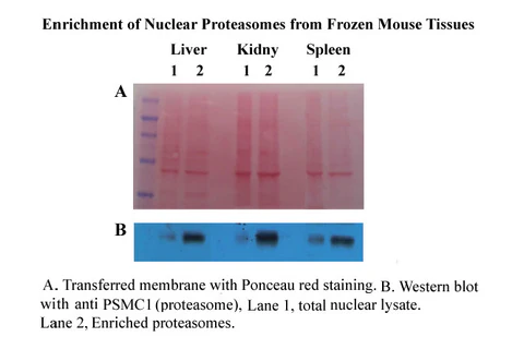
The proteasome is a protein degradation system that requires metabolic energy and polymerization of ubiquitin that is different from lysosome-based protein degradation. The 26S proteasome is made up of two subcomplexes (a 20S proteasome and one or two19S regulatory particles). The proteasomes have been found mainly localized in the cytosol but also detected in the nucleus. Traditionally, ultracentrifugation and affinity isolation are the most common methods for the isolation of proteasomes. These methods, though relatively effective, are usually time-consuming with low yield. Many affinity-based methods require harsh elution conditions that may affect the activity of isolated proteasomes and limit certain downstream applications. In most cases, the methods can’t distinguish proteasomes derived from cytosol or nucleus. To overcome these shortcomings, we have developed spin-column-based kits for the enrichment of proteasome from nuclei (Cytosolic Proteasome Enrichment Kit is also available under Cat # PT-040). The protocol is simple and rapid with a high yield. The gentle protocol maintains the association of proteasomes with ubiquitin and other proteins and is useful for the studies of proteasome structure and function. The highly enriched proteasomes can also be used as a starting material for affinity purifications.
Kit Components:
Items | Quantity |
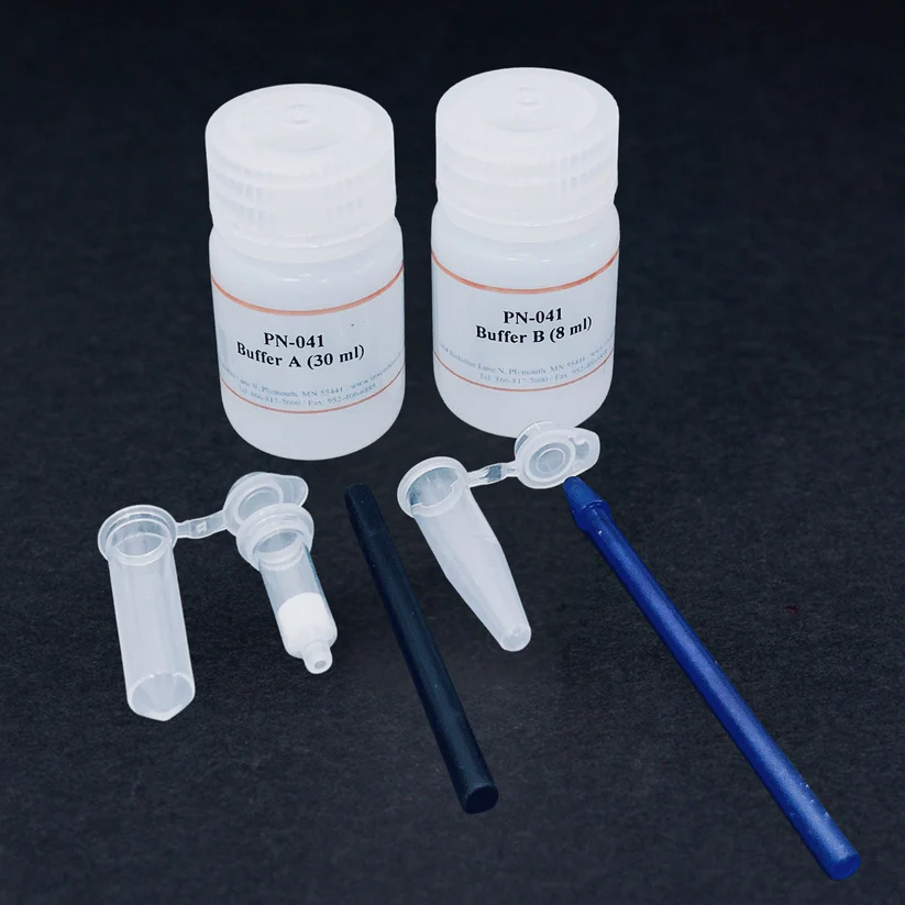
Buffer A | 30 ml |
Buffer B | 8 ml |
Filter Cartridges with collection tube | 40 units |
Pestle for 1.5 ml tube | 2 units |
Protein extraction powder | 2.0 grams |
References (1)
1. Zhang, C. Y., Zhang, R., Zhang, L., Wang, Z. M., Sun, H. Z., Cui, Z. G., & Zheng, H. C. (2023). Regenerating gene 4 promotes chemoresistance of colorectal cancer by affecting lipid droplet synthesis and assembly. World Journal of Gastroenterology, 29(35), 5104-5124.
Minute™ High-Efficiency Saliva Exosome Isolation Kit (50 Preps)
Minute™ High-Efficiency Saliva Exosome Isolation Kit (50 Preps)
Cat# : SE-030
Description
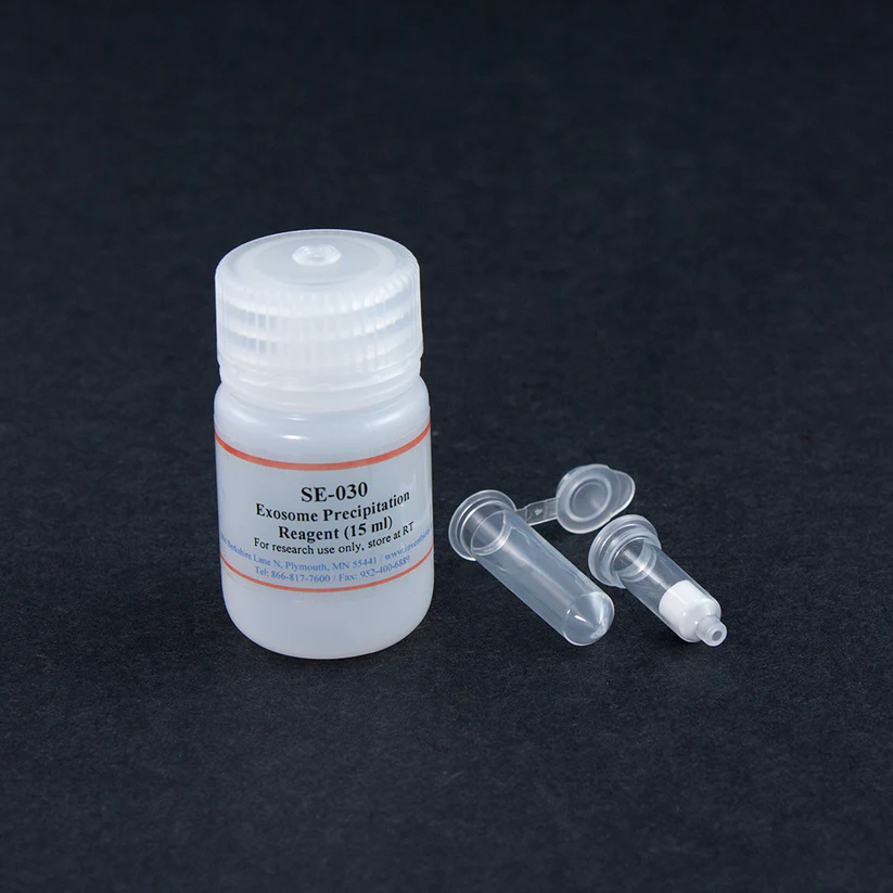
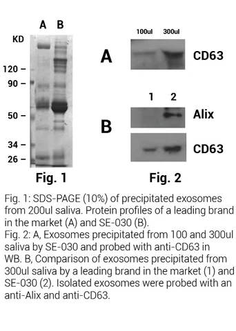
Diagnoses using exosomes derived from saliva have been attracting great attention in recent years due to the ease of sample collection. However, saliva samples are challenging when it comes down to exosome isolation. In addition to cells and cell debris, large amount of amylase, mucin and glycoprotein are present in saliva, and make the samples viscous and hard to manipulate. Pre-treatments, such as sonication and dilution, are often required. In many cases, the saliva samples can still be difficult to handle even after pre-treatments. This product is specifically designed to address these issues using the proprietary saliva filters. Highly viscous saliva samples can be converted to non-viscous solutions instantly by passing the samples through the filter. Exosomes can then be readily precipitated from as little as 100 µl saliva using the highly effective non-PEG-based reagent.
Kit includes:
Items | Quantity |
Exosome Precipitation Reagent | 15 ml |
Filter Cartridge | 50 units |
2.0 ml Collection Tubes | 50 units |
References (2)
- Hyun, K. A., Gwak, H., Lee, J., Kwak, B., & Jung, H. I. Salivary Exosome and Cell-Free DNA for Cancer Detection.
- Jia, Y., Wang, C., Sun, H., & Ni, Z. (2019). Microfluidic Approaches Toward the Isolation and Detection of Exosome Nanovesicles. IEEE Access.
Minute™ Plant Golgi Apparatus Enrichment Kit
Description
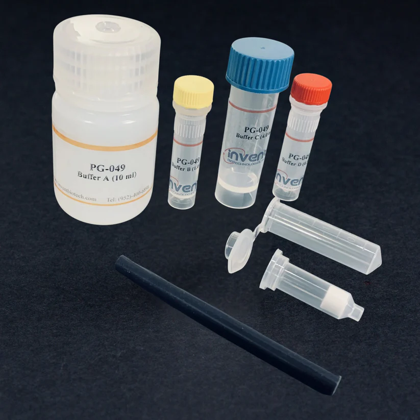
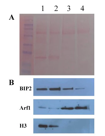
The plant Golgi apparatus plays an important role in the biosynthesis of cellular structural components and protein trafficking. However proteomic characterization of the Golgi has been hindered by limited methodologies for obtaining enriched/purified Golgi fraction. The traditional method for isolating Golgi apparatus depends upon the use of density centrifugation, which is tedious and time-consuming. A large amount of starting material is usually required. Plant Golgi stacks are notoriously labile and loss of stack integrity can occur if harsh homogenization is used. This means that assessment of purity by morphological means such as electron microscopy is difficult. This kit is designed to overcome certain disadvantages of traditional methods by employing spin column-based homogenization, differential centrifugation, and selective precipitation for the enrichment of the Golgi. The protocol is simple and straightforward and requires only a milligram amount of starting material. Significant enrichment of the Golgi fraction (2-3 folds) can be obtained.
Kit includes:
Buffer A | 10 ml |
Buffer B | 1.0 ml |
Buffer C | 4 ml |
Buffer D | 0.5 ml |
Filter Cartridge with Collection Tubes | 20 units |
Plastic Rods | 2 units |
A. Ponceau S stained blot. B. Western blot with ER (BIP2), Golgi (Arf1) and histone 3 (H3) marker antibodies. The antibodies were purchased from Agrisera (Vannas, Sweden). Lanes 1&2: total protein from A. thaliana and N. tabacum; Lanes 3&4: enriched Golgi fractions from A. thaliana and N. tabacum, respectively.
Minute™ Plant Endosome Enrichment Kit
Description
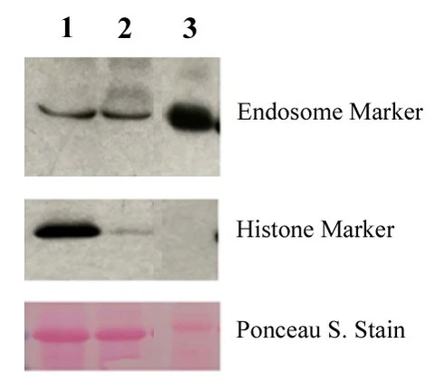
An understanding of endomembrane processes and their biological implication is important for understanding plant growth and development. Endosomes are a heterogeneous collection of organelles in the sorting and delivery of internalized material from cell surface and the transport of materials from Golgi to lysosome or vacuole. In plant cells, two clearly defined endosomal compartments have been identified: the trans-Golgi network (early endosome equivalent) and the multivesicular body (late endosome equivalent). Traditionally, methods for isolation and purification of these two compartments are tedious and time-consuming involving sucrose gradient centrifugation. In some cases, sucrose gradient isolation is coupled with immunoaffinity purification and larger amount of starting materials are usually required (10-50 g). We have developed this kit using a spin-column-based format coupling with selective precipitation for enrichment of endosomal compartment. This approach is easy and rapid and only small amount of starting material is required.
Kit includes:
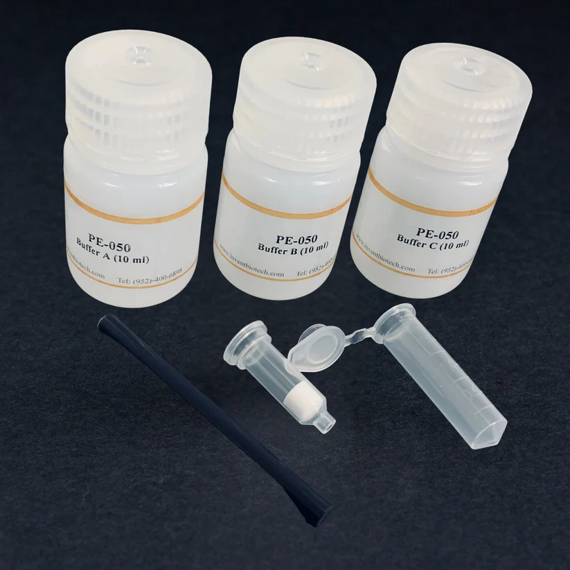
Buffer A | 10 ml |
Buffer B | 10 ml |
Buffer C | 10 ml |
Plastic Rods | 2 units |
Filter Cartridge with Collection Tubes | 20 units |
Detection of Enriched Endosomes from N. benthmiana by Western blotting. The plant leaf that carries GFP-tagged ara6 (early endosome marker) was processed as described in the protocol and probed with anti-ara6 and anti-histone antibodies. Lane 1, Total tissue lysate; lane 2, Supernatant from step 4 of the protocol; lane 3, Enriched endosome fraction.


