Minimizing CRISPR Off-target Effects

Minimizing CRISPR Off-target Effects

Personalized medicine has long been predicted as the ‘future of medicine.’ In fact, with every significant discovery in modern medicine, someone inevitably hails it as the advent of personalized medicine and a revolution in healthcare. Despite this, personalized medicine still seems out of reach for the near future.

The newest avenue with which we aim to reach personalized medicine lies with gene therapeutics. Gene therapies got off to a rocky start in the late ’90s, when several failed clinical trials stalled progress in the field. Despite this, research continued and, starting in 2014, investments, improved molecular tools and novel therapeutics in the field have rebounded and the industry has begun to run with the concept.

CRISPR and Therapeutics

While a few gene therapies have already been approved, there remain some significant problems that the technology faces. While modified viruses remain a popular choice for the delivery of genes, they have primarily proven themselves useful for the replacement of nonfunctional genes 1. The disruption of genes that have gained harmful functions has proven to be more complex but is an ideal use-case for CRISPR.

The CRISPR gene-editing tool came to prominence in 2012 as an efficient method to edit DNA in vivo. It does so by utilizing two main components, the Cas9 endonuclease, and customizable guide RNAs (gRNA(s)), which target the Cas9 to the location where it precisely cuts DNA. Various methods can be used to introduce the CRISPR system into cells, such as transduction by viruses, or direct introduction of the ribonucleoprotein complex (Cas9 & gRNA(s)) by transfection or electroporation. However, commonly these experiments are performed by transfection of high-quality purified recombinant plasmids encoding gene expression cassettes for both the gRNA(s) and Cas9. Learn more about the simplest purification method for CRISPR-ready plasmid DNA here.

Several companies have been exploring the use of CRISPR as the future of gene therapy and the solution to the current problems that exist in the industry. Unfortunately, CRISPR is still a long way off from any human clinical trials. The reason for this, as brought to the forefront by the recent CRISPR baby debacle, is the potential for off-target effects.

Because of the nature of the ribonucleoprotein binding to its target, Cas9–the ‘molecular scissors’ of the complex–can potentially act at a lower efficiency at unintended locations in the genome. The effects of off-target activity range from negligible to initiating transformation of the cell into an early-stage cancer.

Minimizing CRISPR Off-Target Effects

Recently, a paper published by Listgarten et. al. 2 details a combined method of machine learning and efficient genomic searching to minimize the risk of off-target activity by identifying potential off-target sites and predicting their risk of being cut.

These tools, known as Elevation and dsNickFury, allow researchers to use rational design to create their gRNAs, and then predict the off-target effects such a gRNA would have on their system of interest.  This highly accurate modeling program assigns a score to potential off-target sites based on their location in the genome and sequence similarity to the intended target, allowing researchers to quickly determine if a potential off-target should be a source of worry.

The result of these programs assigns and aggregate score to each gRNA, rating it overall in terms of its potential for use in a CRISPR experiment.  By combining these tools with Azimuth, a tool from the same Microsoft team for predicting on-target efficiency, users can gain insight into the complete picture of their gRNA’s expected behavior.

Optimizing for the Future

While we haven’t yet optimized the CRISPR system for human use, tools such as dsNickFury, Elevation, and Azimuth allow users to get one step closer to better design and prediction mechanisms in CRISPR experiments. With technology such as this to aid scientists in predicting and scoring off-target effects in CRISPR systems, we are one step closer to marrying the realms of gene therapy and CRISPR to produce true personalized medicine.

Try the simplest purification method for CRISPR-ready plasmid DNA

Hidden Dangers in Infant Food

Hidden Dangers in Infant Food

When you browse the snack food aisle at your local grocery store or grab a quick bite at a fast-food drive-through, food safety can be easy to take for granted. While many countries have regulatory bodies and food safety practices that make food contamination a rare occurrence, this is not true everywhere. In addition, while food poisoning can be extremely unpleasant, it is typically not life threatening for individuals living in countries with adequate medical care and access to antibiotics. However, these experiences are not universal and many developing countries suffer serious adverse effects from a lack of food safety practices throughout the food processing chain.

Examining Pathogenic Contaminants

Given these issues, Tsai et al.1 set out to examine the diversity of pathogens in infant foods in a low-income area of Kenya in an effort to elucidate if the frequent diarrheal diseases affecting infants in that region were due to pathogenic contaminants. The team collected infant food samples directly from mothers and stored the samples in DNA/RNA Shield to ensure DNA and RNA stabilization during transportation and storage. Once the samples arrived to the laboratory, total DNA and RNA was isolated using the ZymoBIOMICS DNA/RNA Miniprep Kit. After ensuring the DNA and RNA extracts were inhibitor free, the samples were analyzed for the presence of over 35 gene targets indicating the presence of pathogens of interest using the TaqMan® Array Card analysis. For example, the presence of stx1 and stx2 (Shiga toxin production genes) indicated the presence of Shiga toxin-producing E. coli.

The researchers found that 62% of collected food samples were contaminated and that the type and frequency of the contaminants varied by month. Of the foods examined, cow’s milk was deemed to be the most concerning, as it was shown to have a higher likelihood of contamination by enteric pathogens when compared to other common infant foods. These results suggest that exposure to pathogens is constant and high in low-income countries and support the implementation of household water, sanitation, and hygiene interventions in these communities.

Crucial Insight

This research shows how advances in molecular genomics have allowed crucial insight into food contamination and the infections associated with it. Using technology developed by Zymo Research, scientists are able to identify which pathogens are most prevalent in contaminated food and can begin to elucidate how these contaminants are introduced and spread. To learn more about this research and to see how Zymo Research’s products are used in the field, read the paper here.

Try a Free Sample of the ZymoBIOMICS DNA/RNA Miniprep Kit Used in This Study:

References:

1. Tsai, K., Simiyu, S., Mumma, J., Aseyo, R. E., Cumming, O., Dreibelbis, R., et al. (2019). Enteric Pathogen Diversity in Infant Foods in Low-Income Neighborhoods of Kisumu, Kenya. International Journal of Environmental Research and Public Health.
2. https://www.zymoresearch.com


How to Discover Biases in Metagenomic Studies

How to Discover Biases in Metagenomic Studies

The growth of metagenomic studies has revolutionized our understanding of the relationships between microbiota and the environment or health.

While this realization has resulted in many new discoveries, data reproducibility has remained a challenge. This issue spans metagenomic research across labs and stems from the fact that bias can be introduced at various steps across the metagenomics workflow, as observed by many in the field 1-6.

The problem of bias is so widespread that even submitting the same sample to two different microbiome profiling organizations can yield results that are dramatically different from one another (Figure 1).

Figure 1. Inconsistent interpretation of the microbial composition of one stool sample by American Gut and uBiome. The figure was adapted from: “Here’s the Poop on Getting Your Gut Microbiome Analyzed” Science News. 2014.

These biases can arise at every step throughout the entire metagenomics workflow. However, one of the most problematic steps that contributes to bias lies in nucleic acid extraction. With growing evidence of systemic biases, the need for more accurate metagenomic nucleic acid extraction workflows is now larger than ever.

How do Biases Occur Within Extraction?

Microbial communities are complex and diverse, consisting of Gram-positive bacteria, Gram-negative bacteria, and fungi. Accurate metagenome profiling requires the liberation of DNA from all the diverse species within a microbial community. However, it is common to observe ineffective lysis during the nucleic acid extraction which then leads to microbial profile bias. This is due to some microbes being very difficult to lyse 6, 8. If the cells are not lysed, the DNA will remain locked away within the cell and will not be purified or detected.

It has been shown that processes utilizing chemical or thermal lysis overrepresent the easy-to-lyse organisms (Gram-negative bacteria) due to this very reason. Since the tough-to-lyse organisms (e.g. Gram-positive bacteria and yeast) are more resistant to DNA liberation, it causes a bias towards the easy-to-lyse species. Many extraction protocols do not account for these vast differences in sample composition meaning it is common to observe non-uniform lysis and microbial profile bias 9.

Extraction protocols that utilize mechanical lysis (e.g. sonication, blending, liquid nitrogen/mortar and pestle, French pressing, and bead-beating) are considered the best approach to microbial lysis due to their stochastic nature with bead beating referred to as the gold standard. However, these mechanical lysis methods still need to be optimized or they will suffer from issues such as low yield, excessive nucleic acid shearing, non-uniform lysis, excessive heat, and shear forces.

How Can Bias Be Discovered?

The only true way to know if an extraction system is introducing bias into a metagenomic study is to evaluate the system with a microbial standard. A microbial standard refers to a pool of various microorganisms (including both Gram-positive and Gram-negative species) that act as a mock microbial community and mimics the metagenomic populations present within samples. This standard is processed normally through the extraction workflow.

Since the abundance of each microorganism in the microbial standard is known, the results obtained from the 16s sequencing data should match closely to the standard. Large deviations from this indicate that the extraction system introduced bias into the results. Most commonly, these deviations reveal themselves as an overrepresentation of Gram-negative species in the population. This can be seen clearly in a comparison of various extraction systems (Figure 2).

Figure 2. Microbial profiling will under-represent the abundance of hard-to-lyse microbes if the DNA extraction method cannot break open these cells. Four different extraction methods were assessed using the well-defined ZymoBIOMICS® Microbial Community Standard and 16S sequencing.

Bias-free Methods

The ZymoBIOMICS line addresses this key challenge of bias within a metagenomics workflow. The ZymoBIOMICS 96 Magbead DNA Kit utilizes mechanical lysis that has been developed and optimized with microbial community standards to ensure complete lysis of all the tough-to-lyse organisms (Figure 3).

Figure 3: Assessing the performance of four different DNA extraction kits with the ZymoBIOMICS Microbial Community Standard. The four different DNA extraction methods investigated include ZymoBIOMICS 96 DNA Magbead Kit, Human Microbiome Project fecal DNA extraction protocol (HMP Protocol), a soil DNA extraction kit from “Supplier M” and a fecal DNA extraction kit from “Supplier Q”. DNA was extracted with ZymoBIOMICS DNA Miniprep Kit and then subjected to 16S targeted sequencing with an internal library preparation protocol. The microbial composition was determined by mapping raw sequencing reads against reference 16S sequences of the strains contained in the standard. The composition of the purified microbial standard was compared to the theoretical composition and shown to match closely for the ZymoBIOMICS kit which indicates unbiased lysis

Learn more about ZymoBIOMICS 96 MagBead DNA Kit used in this study:

References:
 
  1. Sinha R, Abnet CC, White O, Knight R, Huttenhower C: The microbiome quality control project: baseline study design and future directions. Genome Biol 2015, 16:276.
  2. Hsieh YH, Peterson CM, Raggio A, Keenan MJ, Martin RJ, Ravussin E, Marco ML: Impact of Different Fecal Processing Methods on Assessments of Bacterial Diversity in the Human Intestine. Frontiers in microbiology 2016, 7:1643. 13.
  3. Vishnivetskaya TA, Layton AC, Lau MC, Chauhan A, Cheng KR, Meyers AJ, Murphy JR, Rogers AW, Saarunya GS, Williams DE et al: Commercial DNA extraction kits impact observed microbial community composition in permafrost samples. FEMS microbiology ecology 2014, 87(1):217-230. 14.
  4. Hart ML, Meyer A, Johnson PJ, Ericsson AC: Comparative Evaluation of DNA Extraction Methods from Feces of Multiple Host Species for Downstream Next-Generation Sequencing. PloS one 2015, 10(11):e0143334. 15.
  5. Kennedy NA, Walker AW, Berry SH, Duncan SH, Farquarson FM, Louis P, Thomson JM, Satsangi J, Flint HJ, Parkhill J et al: The impact of different DNA extraction kits and laboratories upon the assessment of human gut microbiota composition by 16S rRNA gene sequencing. PloS one 2014, 9(2):e88982. 16.
  6. Sohrabi M, Nair RG, Samaranayake LP, Zhang L, Zulfiker AH, Ahmetagic A, Good D, Wei MQ: The yield and quality of cellular and bacterial DNA extracts from human oral rinse samples are variably affected by the cell lysis methodology. Journal of microbiological methods 2016, 122:64-72.
  7. Saey TH: Here is the poop on getting your gut microbiome analyzed. In: Science News. vol. 2017; 2014.
  8. Farkaš V, Takeo K, Maceková D, Ohkusu M, Yoshida S, Sipiczki M. Secondary cell wall formation in Cryptococcus neoformans as a rescue mechanism against acid-induced autolysis. FEMS Yeast Research, 2009, 9(2): 311-320
  9. Costea et al. Towards standards for human fecal sample processing in metagenomic studies. Nature Biotechnology(2017) 11:1069-1076
  10. https://www.zymoresearch.com
 

Children of Zika

Children of Zika

As the children born with Zika grow up, more developmental deficits are being noticed. New studies examine these changes from an epigenetic perspective and seek to improve detection of the virus.

But as soon as Zika caught global attention, it disappeared. The World Health Organization declared that Zika was no longer a global emergency and the active outbreaks ended. However, for all the families affected by the virus during that time, their journey is just beginning.

Generation Zika

Zika is a virus spread primarily by mosquitoes and sexual transmission. For most, an infection is quite mild causing minor or no symptoms. However, if an infection occurs during pregnancy, the virus can spread to the baby and result in severe birth defects including microcephaly (an abnormally small head) and brain malformations. Of the mothers with Zika during pregnancy, approximately 6% gave birth to babies with Zika-associated birth defects.

During the epidemic, thousands of babies were born with these birth defects. In Brazil alone, nearly 3000 babies were born with microcephaly. As these babies become toddlers and continue to grow, further developmental delays are beginning to form.

A recent CBS documentary featured the children of “Generation Zika” as they turn 3 years old. The film highlights the array of neurological and physical challenges the children are facing.1 Many toddlers cannot walk or talk and suffer from vision impairment, muscle weakness, and seizures. The only treatment is intensive physical therapy. However, many families come from rural or low socioeconomic backgrounds. This makes paying for care and traveling to frequent appointments infeasible.

Despite the generosity of physical therapists and doctors who have volunteered their time and services, there is simply not enough available care to properly support all the affected children. Currently, the Brazilian government has not provided any financial or medical aid to the families and there are no plans to do so in the future. With the long-term health effects of Zika unclear, the pressing scientific question at hand is whether more Zika related neurological issues will appear later in life for affected children, especially for the 94% of babies who did not exhibit any birth defects.

Examining the Epigenetics

A recent study by Janssens et al. 2 is attempting to address some of the questions that surround the development of babies born to mothers with Zika. Since the Zika virus can pass from a mother to a fetus during pregnancy and directly affect the fetus’s neurological development, researchers examined DNA methylation patterns within the genome of neural cells.

Embryonic stem cell-derived (ESC) brain organoids composed of multiple organized brain tissues were used as the best model for a developing brain. The complex organoida allowed for hetereocellular interactions to occur alongside any resultant regulation of gene expression and epigenetic patterning. 2 The model organoids were then infected with the Zika virus.

Methylation levels were compared using whole-genome bisulfite sequencing to identify any significant methylome changes induced by Zika virus. To do this, researchers utilized the EZ DNA Methylation Gold Kit to perform the bisulfite conversion before constructing libraries.

Changes were identified in the DNA methylation patterns of the infected neurological cells. These changes were found at the genes that have been previously linked to brain disorders. The regions of methylation change were found in excess of 30-40% at regions near transcription start sites. This finding was very significant since the methylation changes near these regulatory regions dramatically influence gene expression, resulting in a variety of physiological symptoms depending on the genes with the epigenetic changes. Using the Direct-zol RNA Miniprep Kit, the group purified RNA and performed qRT-PCR to determine which genes were being expressed.

Advances in Detection

While some researchers have focused on the outlook for the children of Zika, others have focused on optimizing the methods that are used to detect the virus in patients. Typically, diagnosing Zika has relied on detecting Zika RNA or antibodies in serum. But with reports of longer duration of virus shedding at higher concentrations, detection of Zika in urine has been an increasingly popular method. However, detecting Zika in urine possesses unique challenges due to the instability of RNA. Urine provides a good environment for RNase activity, up to 100x higher than in serum.

Due to this, a recent study by S.K. Tan et al. sought to address these issues with Zika stability in urine. The research team evaluated the effect of temperature, initial Zika levels, time between sample collection and extraction, and nucleic acid stabilizers like DNA/RNA Shield.

From their tests, researchers found that urine samples being evaluated for Zika can be stored at room temperature for up to 48 hours without significant impact on the levels of Zika RNA. Storing the urine at 4 °C in this window can help to further minimize degradation. However, if the samples will not be processed within this time frame, the researchers recommended using DNA/RNA Shield to stabilize the specimens before testing. The reagent detected all the Zika virus when compared to cold storage and improved quantitative recovery of the RNA.

In the aftermath of the Zika outbreak, there are many questions still left to answer as we work to improve treatment and detection of the virus. Health officials in several countries will be monitoring the Zika children for years to come to better understand the range of difficulties that they face and see if any problems arise for those children who were mildy affected or were asymptomatic at birth.

Learn more about the Nucleic Acid stabilizer used in this study:

References:

1. Zika: Children of the outbreak. CBSN, 2019. CBSN Originals.
2. Janssens S et. al. Zika Virus Alters DNA Methylation of Neural Genes in an Organoid Model of the Developing Human Brain. ASM. 2018.
3. https://www.zymoresearch.com

Turning Back the Epigenetic Aging Clock

Turning Back the Epigenetic Aging Clock

Aging is a major factor associated with chronic disease, which accounts for nearly two thirds of all deaths and contributes to approximately 75% of annual health care costs in the United States1. As scientists and doctors search for novel therapies and interventions to deal with aging and related diseases, the epigenetic clock has emerged as an important tool able to predict biological, as opposed to chronological, age in mammals2,3. Now, a new study provides the first evidence that reversal of biological age in humans may be possible4.

A 3-Part Treatment Cocktail

As a primary objective of their study, Fahy et al. administered a year-long, 3-part treatment cocktail consisting of recombinant human growth hormone, dehydroepiandrosterone (DHEA), and metformin to restore age-related decline of thymus function in male research subjects ranging from 51-65 years of age5. Using a variety of quantitative and qualitative techniques (MRI, blood cellular composition, cytokine signaling, and inflammation), the authors reported strong signs for reversal of the thymic damage and declining immune function often associated with age.

Turning Back the Clock

The authors next measured the biological age of each study participant using the epigenetic clock – identifying it as a simple yet compelling way to assess systemic aging. In agreement with the immunological and thymic measurements, Fahy et al. found an average reduction of 2.5 years of biological age in the research subjects. Furthermore, the age reversal seemed to accelerate the longer the recipients received treatment. Importantly, the age-related therapeutic benefits persisted up to 6 months after treatment concluded.

Whereas earlier studies showed that the epigenetic clock could be slowed down through various lifestyle or environmental modifications6, the study by Fahy et al. represents the first evidence that biological age is reversible. Commenting on their work, the authors noted that the epigenetic clock “…is the most accurate measure of biological age and age‐related disease risk available today. This justifies the use of epigenetic clocks to estimate the effectiveness of putative aging interventions on a practical timescale.”

We now offer DNAge Services, a Next-Gen Sequencing based platform to analyze the biological age of human and mouse DNA samples. Zymo Research’s technology expands upon the original epigenetic clock by utilizing bisulfite sequencing and a unique capture strategy to target a panel of DNA methylation biomarkers that are highly informative of aging. The DNAge Epigenetic Clock Service can be applied to not only anti-ageing intervention studies but also investigation of age-related diseases.

Learn More about the DNAge Services:

References:


1. Raghupathi, W. , and Raghupathi, V. An empirical study of chronic diseases in the United States: A visual analytics approach to public health. Int J Environ Pres Public Health. 15(3):431 (2018).
2. Horvath, S. DNA methylation age of human tissues and cell types. Genome Biol. 14, R115 (2013).
3. Field, A. E. et al. DNA methylation clocks in aging: categories, causes, and consequences. Mol. Cell 71, 882–895 (2018).
4. Abbott, A. First hint that body’s ‘biological age’ can be reversed. Nature 573, 173 (2019).
5. Fahy, G.M. et al. Reversal of epigenetic aging and immunosenescent trends in humans, Aging Cell (2019). DOI: 10.1111/acel.13028
6. Quach, A. et al. Epigenetic clock analysis of diet, exercise, education, and lifestyle factors. Aging 9, 419–446 (2017).
7. https://www.zymoresearch.com

One Single Cell At A Time

One Single Cell At A Time

Multicellular development begins with a single cell

Each person on the planet is made up of over 30 trillion cells — all originating from a single cell. Those cells make up hundreds of different cell types, with distinct roles ranging from protection against infections, to carrying messages from extremities to our brain, which are essential for life. A long-standing goal in biological science is to understand how each individual cell is specified and how it contributes to the whole of an organism during development, and what goes wrong during disease onset. In just a few centuries, researchers have gone from barely being able to visualize a cell, to describing virtually every genetic molecule within a cell attributed to the advent of Next-Gen Sequencing (NGS).

To further accomplish this goal, large international collaborative projects like the Human Cell Atlas (HCA), Chan Zuckerberg Initiative (CZI), and Human BioMolecular Atlas Program (HuBMAP) have been created with the goal of using single-cell NGS techniques to identify and describe every cell type in the human body — a single cell at a time.

Single-cell methylome

At the forefront of using Next-Gen Sequencing technologies is the ability to investigate the epigenome. One of the most well studied and fundamental epigenome marks, DNA methylation, has been demonstrated to be vital for essential cellular processes including imprinting and cell-type-specific regulation of gene expression.

Current mainstream methodologies, such as whole-genome bisulfite sequencing (WGBS-seq), and array-based techniques query DNA methylation using DNA collected from thousands to hundreds of thousands of cells (bulk sequencing). By isolating DNA from thousands of cells at a time, it is possible to miss the heterogeneity present and mask signals from rare cell populations like stem and cancer cells. Recently, bisulfite sequencing for single-cell applications has been developed, allowing for unprecedented exploration of methylation heterogeneity between cells, as well as the identification of new cell types and regulatory networks that distinguish the newfound cell types1–5.

In a recent Science paper, researchers from Professor Joseph Ecker’s lab at the Salk Institute developed a protocol (snmC-seq) to isolate single nuclei from the frontal cortex of mice and human tissue samples. Cells were sorted into wells of a 384-well plate, and DNA within single nuclei was bisulfite converted using Zymo EZ DNA Methylation-Direct chemistry, pooled, and resulting libraries sequenced. They were able to define methylation signatures for 16 and 21 cell subpopulations in mice and human samples respectively, including a new cell type5.

Another new method, sci-MET5 was developed utilizing Zymo EZ DNA methylation technology. Taking advantage of these exciting technologies, mapping DNA methylation at single-cell levels revealed extensive heterogeneity within the methylomes of diverse cell types including oocytes, fibroblasts, liver and brain3–8. With normal single-cell methylomes of diverse cell types sequenced, future researchers can now start to study single-nuclei methylomes in the context of disease states to investigate cell-type-specific changes and discover new biomarkers.

New Frontiers

The level of methylation at specific locations and the overall three-dimensional (3D) architecture of chromosomes is known to regulate gene expression, but how they each regulate the other is not clear. Now, with the advent of single-cell multi-omics techniques DNA methylation, chromatin accessibility, transcription, and 3D chromosomal structures, in various combinations, can be measured from the same cell simultaneouslyl9–13. For example, two methods, scM&T-seq9 and scNMT-seq10 were used to uncover new relationships between chromatin accessibility, methylation, and transcription in stem cells. Using these techniques, novel relationships between the dynamics of methylation and transcriptions and how these relationships regulate developmental trajectories are starting to be revealed.

In order to investigate 3D chromatin architecture and methylation in the same cell, two new methods were developed. scMethyl-HiC11 was used to examine stem cells and sn-m3C-seq12 was used to investigate brain cells. Both groups of researchers demonstrated the presence of cell-type-specific architectures associated with specific methylation patterns. Measuring these events simultaneously has revealed valuable insights into how DNA methylation, chromatin accessibility, and gene expression are regulated. The integration of multiple layers of ‘omics’ information allows for the relationships between methylation, 3D architecture, and transcription to be elucidated.

Single-cell DNA methylation sequencing technologies are changing researchers’ understanding of cell type diversity within entire organisms and allowing unparalleled resolution of developmental and disease onset events. Zymo Research is proud to contribute to this new revolution and continues to support innovation and ground-breaking research.

Learn more about Zymo EZ methylation technology used in this blog:

References:

1. Smallwood, S. A. et al. Single-cell genome-wide bisulfite sequencing for assessing epigenetic heterogeneity. Nat. Methods 11, 817–820 (2014).
2. Farlik, M. et al. Single-Cell DNA Methylome Sequencing and Bioinformatic Inference of Epigenomic Cell-State Dynamics. Cell Rep. 10, 1386–1397 (2015).
3. Gravina, S., Ganapathi, S. & Vijg, J. Single-cell, locus-specific bisulfite sequencing (SLBS) for direct detection of epimutations in DNA methylation patterns. Nucleic Acids Res. 43, e93 (2015).
4. Mulqueen, R. M. et al. Highly scalable generation of DNA methylation profiles in single cells. Nat. Biotechnol. 36, 428–431 (2018).
5. Luo, C. et al. Single-cell methylomes identify neuronal subtypes and regulatory elements in mammalian cortex. Science 357, 600–604 (2017).
6. Gravina, S., Dong, X., Yu, B. & Vijg, J. Single-cell genome-wide bisulfite sequencing uncovers extensive heterogeneity in the mouse liver methylome. Genome Biol. 17, (2016).
7. Yu, B. et al. Genome-wide, Single-Cell DNA Methylomics Reveals Increased Non-CpG Methylation during Human Oocyte Maturation. Stem Cell Rep. 9, 397–407 (2017).
8. Wei, Y. et al. DNA methylation analysis and editing in single mammalian oocytes. Proc. Natl. Acad. Sci. 116, 9883–9892 (2019).
9. Angermueller, C. et al. Parallel single-cell sequencing links transcriptional and epigenetic heterogeneity. Nat. Methods 13, 229–232 (2016).
10. Clark, S. J. et al. scNMT-seq enables joint profiling of chromatin accessibility DNA methylation and transcription in single cells. Nat. Commun. 9, (2018).
11. Li, G. et al. Joint profiling of DNA methylation and chromatin architecture in single cells. Nat. Methods 1–3 (2019) doi:10.1038/s41592-019-0502-z.
12. Lee, D.-S. et al. Simultaneous profiling of 3D genome structure and DNA methylation in single human cells. Nat. Methods 1–8 (2019) doi:10.1038/s41592-019-0547-z.
13. Pott, S. Simultaneous measurement of chromatin accessibility, DNA methylation, and nucleosome phasing in single cells. eLife, e23203 (2017).
14. https://www.zymoresearch.com


Can Liquid Biopsies Enable Early Detection of Renal Cancer?

Can Liquid Biopsies Enable Early Detection of Renal Cancer?

Renal cancer remains among one of the most lethal genitourinary cancers, and is one of the top 10 most diagnosed cancers overall 1. The incidence of renal cancer has been increasing over the past few decades, puzzling doctors and researchers alike without explanation [1]. Renal cancers are often asymptomatic until progressed to an advanced state, making it a particularly difficult challenge. Given these factors, diagnosis at early stages of disease will be highly valuable in the development of treatment regimens. However, early detection of the renal cancer still requires much investigation.

A Method With Potential

Many researchers are currently working to identify early-stage biomarkers of renal cancer. One example of many promising works was described in an article by Skrypkina et al. in Disease Markers 2. The authors used cell-free DNA as a source of biomarkers for renal cancer detection. They specifically evaluated methylation profiles of circulating cell-free DNA in blood to determine if there is a distinguishable methylation pattern between renal cancer patients and healthy control cohort. The authors discovered that various gene fragments detected in the cell-free DNA of cancer patients were hypermethylated, demonstrating the potential of this method to serve as an early diagnosis tool for renal cancer.

The study began by isolating the plasma from blood extracted from patients with renal tumors then isolated the DNA. Quantification was performed using the Human HCT116 DKO Nonmethylated DNA standards. Next, the EZ DNA Methylation Kit was used to bisulfite convert the isolated DNA in preparation for bisulfite sequencing analysis to identify the methylation marks. The study showed that there was a measurable increase in the amount of cell-free DNA in the blood of patients with renal cancer and was hypermethylated at several key sites. The authors posit that these observations identify key potential markers important for development of early diagnosis tools for renal cancer.

The Future

Their results indicate that a combination of genetic differences was responsible for the observed phenotype. One surprising result was that the majority of genomic effectors were derived from the mitochondrial genome, and this included important genes such as COX1 (Cytochrome c oxidase subunit 1) which created a significant heat tolerance phenotype in the hybrid yeast. Genetic screening and similar genomic techniques, made possible by the technological revolution in genomics, are allowing greater insight into molecular genetics. These studies are crucial in establishing methods for us to evaluate the process of speciation on a molecular level. To read more about this research, see the paper here.

Learn more about the EZ DNA Methylation Kits that were used in this blog:

References:


[1] American Cancer Society. (2018, January 4). Key Statistics About Kidney Cancer. Retrieved February 11, 2019, from Cancer.org:
https://www.cancer.org/cancer/kidney-cancer/about/key-statistics.html


[2]Skrypkina, I., Tsyba, L., Onyshchenko, K., Morderer, D., Kashparova, O., Nikolaienko, O., et al. (2016). Concentration and Methylation of Cell-Free DNA from Blood Plasma as Diagnostic Markers of Renal Cancer. Disease Markers, 3693096.

[3] https://www.zymoresearch.com

Viral Targets: What Makes A Good COVID-19 RT-PCR Test?

Viral Targets: What Makes A Good COVID-19 RT-PCR Test?

Target Selection Tips for SARS-CoV-2 Tests

Since the initial discovery of COVID-19 outbreaks in Wuhan, China over a year ago, the market for diagnostic testing has grown tremendously. Because of their high sensitivity, real-time reverse transcription polymerase chain reaction (rRT-PCR) tests that use fluorescent signals to detect SARS-CoV-2 RNA targets continue to dominate the global testing market. With over a hundred rRT-PCR tests for COVID-19 currently authorized by the FDA, it can be challenging to determine which tests have the best performance.

Detection Details 

SARS-CoV-2 targets commonly detected by rRT-PCR tests include the nucleocapsid (N), envelope (E), spike protein (S), RNA‐dependent RNA‐polymerase (RdRp, also known as nsp12), and open reading frame1ab (ORF1ab) genes 1. N, E, and S are structural proteins while RdRp and ORF1ab are important for the replication of viral RNA. The N protein packages viral RNA into a helical nucleocapsid, the E protein forms a structure that encloses viral RNA, and the S protein consists of glycoprotein spikes that facilitate the invasion of host cells


An optimal COVID-19 rRT-PCR test would detect targets that are highly specific for SARS-CoV-2 and therefore do not exhibit cross-reactivity with other organisms (including other viruses and coronaviruses). This would minimize the chance of false positive results. Target regions should also have a low rate of mutation to maintain the assay sensitivity as new strains of the virus emerge over time. The target region should also be suitable for designing efficient primers and probes.

The nucleocapsid (N) protein of SARS-CoV-2 has 90% amino acid homology with SARS-CoV (SARS, the illness that swept through Asia in 2003), which indicates that this gene is stable and that the sequence has been conserved over time 3. Primers and probes designed on the divergent regions of the N gene will ensure the specific detection of SARS-CoV-2, making the N gene overall one of the most promising targets for SARS-CoV-2 detection. Contrary to the N gene, point mutations in the RdRp gene have been well documented due to the ability of these mutations to interfere with both diagnostic assays and anti-viral treatments such as Remdesivir 4. Similarly, multiple mutations in the S gene have been identified in three new SARS-CoV-2 variants that have emerged in the UK, South Africa, and Japan within the last two months 5. Mutations in the S gene are more likely to occur because they confer a genetic advantage to the virus in terms of increased transmissibility 6.

Single Viral Target vs. Multi-Viral Target TEST

There is common perception that rRT-PCR tests detecting multiple SARS-CoV-2 gene targets perform better than those using a single SARS-CoV-2 target. However, it is important to understand that the number of targets detected by an assay is not an indication of test sensitivity or performance. In fact, a study published in Nature Biotechnology compared over 150 rRT-PCR tests authorized by the American FDA and found that over 25% of the tests were designed using a single viral target 7. In the same study, the top six tests with the lowest limit of detection (highest sensitivity) used exactly one SARS-CoV-2 target 7. This indicates that detecting a single SARS-CoV-2 target does not reduce test performance, as many of the most sensitive tests on the market actually only detect one viral target.

One reason behind the common misconception that it is necessary to have multiple SARS-CoV-2 targets for rRT-PCR is the concern that the virus could mutate in a binding site for the primer and/or probe. This event would ultimately invalidate a single viral target test, but not a multi-target test. Although possible, this is very unlikely to happen, particularly if the selected single viral target is classified as “stable.”

This is particularly true for coronaviruses. This virus family possesses genomes of 26 to 32 kb in length, which is larger than many other RNA viruses (in comparison, HIV and Influenza A genomes are 9.2 and 13.6 kb in length, respectively). Viruses with larger genomes experience higher fitness costs associated with replication errors and mutations8. To compensate for this, coronaviruses (unlike most virus families) use the nonstructural protein 14 (nsp14) to remove misincorporated ribonucleotides before a new RNA strand is extended 8.

The result is that coronaviruses make far fewer replication errors than other RNA viruses, and therefore have fewer mutations and less genetic variability over time 8. For example, it has been estimated that SARS-CoV-2 mutates at a rate that is approximately one-half that of influenza and one-quarter that of HIV 9. Because SARS-CoV-2 is mutating much slower than other RNA viruses, it is not as critical to develop diagnostic testing that prepares for the chance of mutation.

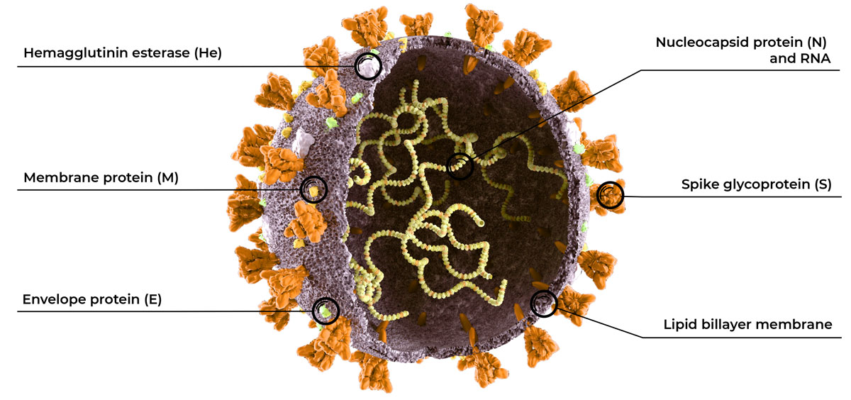
Furthermore, in order to invalidate a single-target rRT-PCR test, a mutant viral strain would need to become established among the population. This is only possible if the mutation confers an evolutionary advantage to the virus. An example is the new SARS-CoV-2 variant that was first detected in the UK in December 2020 and is now spreading worldwide 6. This SARS-CoV-2 variant has multiple mutations in the S gene (which encodes the spike protein) that increase the transmissibility of the strain, which is a clear evolutionary advantage. Most mutations, however, are deleterious or neutral, meaning their frequency in the population will remain negligible and they will not compromise the performance of single-target rRT-PCR tests, especially those designed on a genetically stable region like certain parts of the N gene.

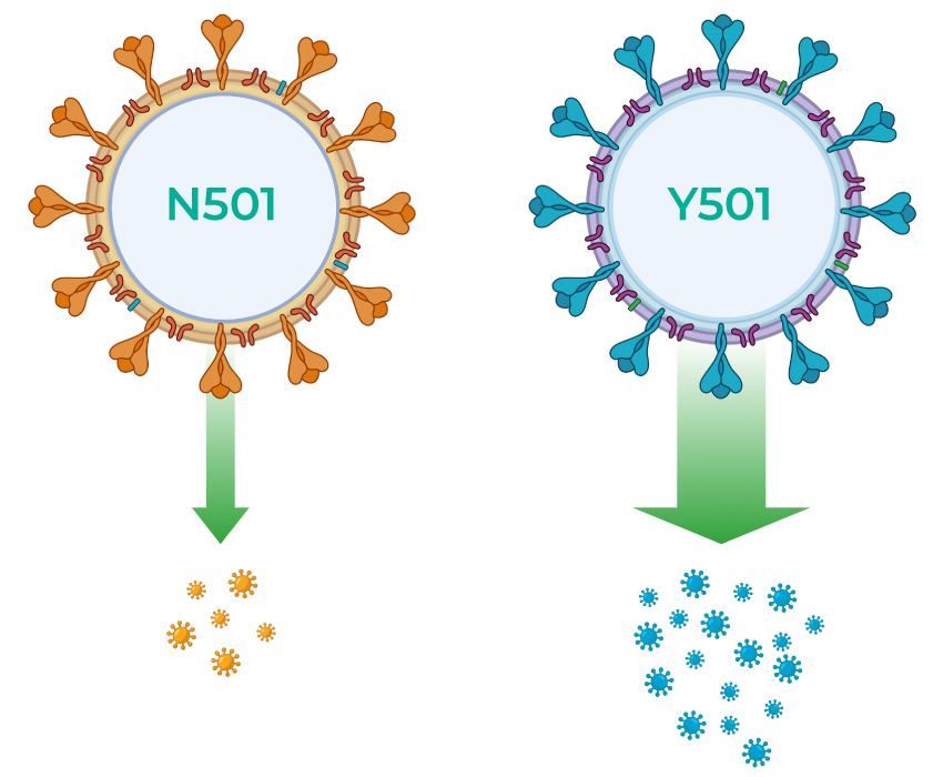
FIG: The variant strain of SARS-CoV-2 that first became prevalent in the UK contains multiple mutations in the spike (S) protein, one of which is an amino acid change from asparagine (N) to tyrosine (Y) at position 501 in the receptor-binding domain of the protein10. The spike protein is under strong selective pressure because it directly affects the transmissibility of the virus.

The Challenges

Detecting multiple targets during rRT-PCR can also increase the risk of having technical difficulties and obtaining ambiguous results. When more targets are included in the same multiplex rRT-PCR test, the reaction becomes more complex and the chance of technical issues (e.g. the formation of unspecific products during the reverse transcription and/or PCR stages) increases. This can dramatically reduce the test performance in cases where the assay design has not been carefully optimized or the testing protocol is altered in any way (e.g. the adoption of a different sample type or different laboratory instrument compared to those recommended by the test manufacturer).

Additionally, it can prove to be more challenging to interpret results from a test that uses multiple targets rather than a single target. In the case that one target amplifies, and another does not (which can happen when the viral load in a sample is approaching the limit of detection of the test), it is often necessary to re-test the patient before an accurate result can be reported 11.

Choosing a Test

It is important to select a test that includes a genetically stable viral target (such as certain regions of the N gene) and is highly specific for SARS-CoV-2. Whether these criteria are met with one or multiple SARS-CoV-2 targets is ultimately a matter of preference, and this does not directly correlate to the overall performance of the test. The limit of detection is the best measurement of test performance, and this should be weighed along with the pricing and sample throughput potential when choosing a COVID-19 testing option.

Zymo Research’s Quick SARS-CoV-2 Multiplex Kit is the newest COVID-19 testing solution to achieve CE-IVD marking, and it has been submitted to the FDA for pre-EUA review. Each sample is analyzed in a single reaction to detect both SARS-CoV-2 and a human internal control during rRT-PCR, yielding accurate results in less than 2 hours. The test has a very low limit of detection of 167 viral copies/ml of sample (10 viral copies/reaction), making it one of the most sensitive assays on the market. The kit is also compatible with high-throughput platforms and is offered at a very competitive price.

References:

1. Li C, Zhao C, Bao J, et al. Laboratory diagnosis of coronavirus disease-2019 (COVID-19). Clinica Chimica Acta, 510:35-46. Published 2020 Nov. doi:10.1016/j.cca.2020.06.045.
2. Significance and Difference between Target Regions for SARS-CoV-2. PerkinElmer Applied Genomics. Published 2020 Apr 12. https://perkinelmer-appliedgenomics.com/2020/04/06/significance-and-difference-between-target-regions-for-sars-cov-2/
3. Dutta NK, Mazumdar K, Gordy JT. The Nucleocapsid Protein of SARS-CoV-2: a Target for Vaccine Development. Journal of Virology, 94(13):e00647-20. Published 2020 Jun 16. doi:10.1128/JVI.00647-20.
4. Martinot M, Jary A, Fafi-Kremer S, et al. Remdesivir failure with SARS-CoV-2 RNA-dependent RNA-polymerase mutation in a B-cell immunodeficient patient with protracted Covid-19. Clinical Infectious Diseases, ciaa1474. Published 2020 Sept 28. DOI: https://doi.org/10.1093/cid/ciaa1474
5. Meredith S. Japan has found a new Covid variant. Here’s how it compares to virus strains in the UK, South Africa. CNBC. Published 2021 Jan 11. https://www.cnbc.com/2021/01/11/japan-covid-variant-how-it-compares-to-strains-in-uk-south-africa.html
6. Rapid increase of a SARS-CoV-2 variant with multiple spike protein mutations observed in the United Kingdom. European Centre for Disease Prevention and Control, Published 2020 Dec 20. https://www.ecdc.europa.eu/sites/default/files/documents/SARS-CoV-2-variant-multiple-spike-protein-mutations-United-Kingdom.pdf
7. MacKay MJ, Hooker AC, Afshinnekoo E, et al. The COVID-19 XPRIZE and the need for scalable, fast, and widespread testing. Nature Biotechnology 38, 1021–1024. Published 2020 Sept. https://doi.org/10.1038/s41587-020-0655-4
8. Rausch JW, Capoferri AA, Katusiime MG, et al. Low genetic diversity may be an Achilles heel of SARS-CoV-2. Proceedings of the National Academy of Sciences. Published 2020 Oct 6. https://www.pnas.org/content/117/40/24614.short
9. Callaway E. The coronavirus is mutating – does it matter? Nature News. Published 2020 Sept 8. https://www.nature.com/articles/d41586-020-02544-6
10. Interim: Implications of the Emerging SARS-CoV-2 Variant VOC 202012/01. Centers for Disease Control and Prevention. Published 2020 Dec 29. https://www.cdc.gov/coronavirus/2019-ncov/more/scientific-brief-emerging-variant.html
11. Giri B, Pandey S, Shrestha R, et al. Review of analytical performance of COVID-19 detection methods. Analytical and Bioanalytical Chemistry. Published 2020 Sept 18. https://link.springer.com/article/10.1007/s00216-020-02889-x
12. https://www.zymoresearch.com

Jan 19, 2021

Biolinkk is now an authorized distributor of CellMosaic®, Inc

Biolinkk is now an authorized distributor of CellMosaic®, Inc.

In January, 2021 , we have collaborated with a new company CellMosaic®, Inc. based in Woburn, Massachusetts, (US). CellMosaic®, Inc. is an innovator and leader in bioconjugation and crosslinking technologies. The company was established in 2008 by Dr. Huang, a technological entrepreneur with profound interest and experience in the conjugation field. Dr. Huang has a PhD in Chemistry from CWRU in Cleveland and received postdoctoral training in biology under Nobel Laureate H. Gobind Khorana from MIT. 

CellMosaic® has spent a lot of research effort on developing advanced conjugation processes that involve controlled, site-specific, or single labeling methods. These processes can generate single ratio conjugates with greater than 90% purity. They have built a successful custom conjugation service business based on these processes and delivered more than 150 projects and 100 unique conjugates. Their customers covering a variety of compounds, including proteins, antibodies, peptides, oligos, enzymes, and chemotherapeutic agents. Most of the conjugation methods and technique know-how are protected as trade secrets.

Products and Services offered by CellMosaic

Biolinkk is now an authorized distributor of Canvax Biotech S.L.

Biolinkk is now an authorized distributor of Canvax Biotech S.L.

We have recently collaborated with a new company Canvax Biotech S.L. based in Córdoba (Spain). Canvax is a worldwide leading Expert in Molecular Biology and GPCR Expression in Heterologous Cells. Canvax is Original Manufacturer & Supplier of the Most Innovative Solutions, Services, Kits and R&D Reagents inside the Molecular & Cell Biology field and has focused on R&D of Multiplex High Throughput Platforms (HTS) for Drug discovery and Diagnostic applied Biosensors.